AMH Ovarian Reserve Calculator: Anti-Mullerian Hormone by Age
Convert your AMH between ng/mL and pmol/L, adjust for the assay your laboratory used, and see where your value sits against published age-stratified reference data.
In short: Convert your AMH between ng/mL and pmol/L, adjust for the assay your laboratory used, and see where your value sits against published age-stratified reference data. Use the calculator above, then read the guide below to interpret your result and its limitations.
The calculator
What AMH is and what it reflects
Anti-Mullerian hormone is a glycoprotein in the transforming growth factor beta family. In women it is produced by the granulosa cells of preantral and small antral follicles, the growing follicles that have left the dormant primordial pool but have not yet been selected for ovulation. Because the number of these small growing follicles mirrors the size of the remaining primordial pool, the AMH concentration in blood serves as an indirect read-out of the ovarian reserve: the stock of follicles a woman still carries. It was first described for its role in male fetal development, where it causes the Mullerian ducts to regress, and its reproductive use in women came later, once sensitive immunoassays made routine measurement practical.
Several practical properties made AMH the most used ovarian reserve marker. Unlike follicle stimulating hormone (FSH), it shows little variation across the menstrual cycle and between cycles in the same woman, so the blood sample can be drawn on any cycle day. It also tends to fall earlier than FSH rises when the reserve begins to shrink, which is why clinicians consider it the more sensitive early signal of ovarian aging. The measurement itself is a two-site sandwich immunoassay, available in manual ELISA formats and on automated platforms.
What AMH measures, and what it does not
This distinction matters more than any number on this page. AMH measures the quantity of the remaining follicle pool, not its quality. It predicts, with good accuracy, how the ovaries will respond to the gonadotropin medication used in IVF: a low AMH flags a likely poor response with few eggs retrieved, while a high AMH flags a likely strong response and a higher risk of ovarian hyperstimulation syndrome. Fertility clinics use it to individualize stimulation doses, which is its best validated clinical job.
What AMH does not do is predict natural fertility. In the Time to Conceive study published in JAMA in 2017, researchers followed women aged 30 to 44 who were trying to conceive without fertility treatment, and found that low AMH (or high FSH) was not associated with a reduced chance of conceiving in a given cycle. Women with AMH in the lowest quarter conceived at similar rates to women with normal values. Professional guidance says the same: the American College of Obstetricians and Gynecologists holds that ovarian reserve testing cannot be extrapolated to predict the likelihood of spontaneous conception, and the ASRM Practice Committee describes these tests as predictors of stimulation response, not of natural fertility. If you are trying to conceive without treatment, age remains by far the strongest predictor of your chance of pregnancy, and a low AMH result should not be read as a verdict that natural conception is impossible.
Related limits follow. AMH says nothing about egg quality, which is driven mainly by age and determines the chance that an embryo is chromosomally normal. It cannot diagnose infertility on its own, it cannot time intercourse, and it is not a screening test that healthy young women need routinely. Its sensible uses are the ones the evidence supports: estimating likely response to ovarian stimulation, helping time discussions about fertility preservation before gonadotoxic treatment, contributing to the assessment of diminished ovarian reserve in women over 35 who have not conceived, and giving a rough indication of where a woman sits on the road toward menopause.
How this calculator interprets your result
The calculator does three things in sequence. First, it converts your value between ng/mL and pmol/L using the exact factor stated in the Roche Elecsys method sheet: multiply ng/mL by 7.14 to get pmol/L, or divide pmol/L by 7.14 to get ng/mL. Second, it harmonizes your value to the assay scale used by the reference data. The reference model behind this page was built on the Immunotech/Beckman IBC scale, and the Gen II assay uses the same standards, so Gen II values are used as entered. Access AMH values are converted with the published equation Access = 0.128 + 0.781 x Gen II, and Elecsys values with Elecsys = 0.087 + 0.729 x Gen II, each inverted to recover the Gen II equivalent. These are regression approximations from assay-comparison studies, not exact translations, which is why the result panel repeats the assay warning every time.
Third, it places your harmonized value against published age-stratified reference data. The age-group medians come from Cui and colleagues, who reported median AMH of 2.35 ng/mL for ages 20 to 31, 1.58 for 32 to 34, 1.30 for 35 to 37, 0.96 for 38 to 40, 1.05 for 41 to 43, and 0.67 above age 43, measured in Chinese women. The spread around those medians comes from the validated model of Kelsey and colleagues (PLoS One 2011), whose residual standard deviation is 0.263 log10 units, giving the 68 percent and 95 percent prediction bands shown in the chart. Your estimated percentile is computed on that log scale. Call it an informed estimate rather than a laboratory percentile: it combines a published median with a published spread, and your own laboratory's reference range, derived from its own population and assay, always takes precedence.
One technical honesty note: the published model works on the log10(AMH + 1) scale, which compresses differences near zero. A value of zero therefore lands at a low but finite estimated percentile rather than at the absolute bottom of the distribution. That is a property of the published mathematics, not a judgment that zero is "not that low": an undetectable AMH means the measurable reserve is exhausted, whatever percentile the curve assigns.
The band labels are deliberately plain. "Within the expected range" (25th to 75th estimated percentile) means your value sits where the middle half of the reference population sits. "Below average" or "low" means your value is below most women your age in the reference data, which is the pattern seen in diminished ovarian reserve, while "high" or "very high" means it sits above most, the pattern seen with polycystic ovary morphology. None of these labels is a diagnosis. They describe where a number falls in a reference distribution, and the clinical meaning always depends on why the test was ordered.
AMH and age: the decline in context
AMH is low at birth, rises through childhood, and peaks in the mid-twenties. In the validated model of Kelsey and colleagues, built from 3,260 data points in healthy females from before birth to the menopause, the peak falls at an average age of 24.5 years. From there it declines for the rest of reproductive life and becomes undetectable around the time of menopause. The model also carries a humbling statistic: age explains only about 34 percent of the variation in AMH. The remaining two thirds come from everything else, genes, body weight, smoking, prior ovarian surgery, hormonal contraception and plain individual difference, which is why two healthy women of the same age can have very different values and both be normal.
The large US study of Seifer and colleagues gives the decline a concrete pace. Across 17,120 women aged 24 to 50 presenting to fertility centers, the median AMH fell by an average of 0.2 ng/mL per year up to age 35, then by 0.1 ng/mL per year thereafter. Those are median rates in a fertility-clinic population, not a rule for any individual, but they explain why clinicians interpret the same number very differently at 28 and at 42: an AMH of 1.2 ng/mL sits comfortably in the middle of the distribution at 40 and near the bottom of it at 28. The Seifer authors themselves warned that their age-specific values cannot be used in isolation to counsel a woman about her chance of having a child; they are one component of an evaluation, not a fertility forecast.
Because AMH tracks the shrinking follicle pool, researchers have asked whether it can predict the age of menopause. The honest answer is: roughly, and only in population terms. In a Dutch model, a 30-year-old with an AMH near 0.15 ng/mL had a predicted median menopause age of 48.8 years, while one with an AMH near 4.38 ng/mL had a predicted median of 55.3 years. That is a real association, and AMH does add information beyond age alone, but the prediction bands for any individual woman are wide, the models are assay dependent, and no blood test can hand you a personal menopause calendar. Treat menopause prediction as a rough indicator of reproductive lifespan, not a date.
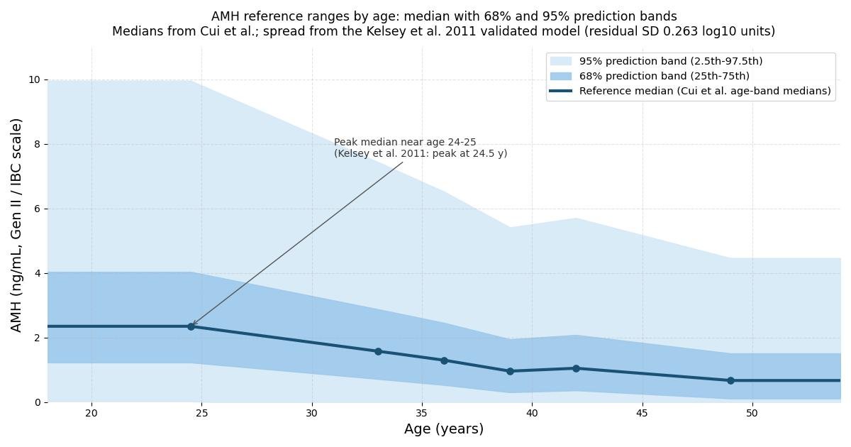
Reference medians by age band (Cui et al.) with 68% and 95% prediction bands from the published residual spread of the Kelsey et al. 2011 validated model. Your result is compared against this reference on the Gen II assay scale.
Assay differences: why the same blood can give different numbers
This section deserves emphasis because it is the most common source of confusion and anxiety. There is no single AMH assay. Laboratories use the manual Beckman Coulter Gen II ELISA, the automated Beckman Coulter Access AMH, the Roche Elecsys (cobas) assay, the Ansh Labs picoAMH ultrasensitive assay, and others. They use different antibodies, different calibrators and different detection chemistries, and their numbers are not interchangeable. In head-to-head comparisons, the Elecsys assay reads roughly 20 to 30 percent lower than the Gen II assay on the same samples (published slopes of about 0.73 to 0.81 with correlations near 0.98), and the Access assay reads lower than Gen II as well (slope about 0.78). Correlations between assays range from about 0.81 to 0.94 depending on the pair and the AMH level, and the agreement is level dependent, meaning the gap between assays is not a constant you can memorize.
The practical consequences are straightforward. Never compare an AMH from one laboratory with a reference range from another, and never track your AMH over time across laboratories that use different platforms. If you repeat the test, use the same laboratory and the same assay. When a clinician or a calculator converts between assays, as this one does with the published regression equations, treat the converted number as an approximation with an error margin, not as the value the other assay would have reported. Standardization efforts continue, but at the time of writing there is still no universal AMH standard that makes all assays agree.
High AMH: PCOS and stimulation response
A high AMH most often means a large antral follicle pool. In polycystic ovary syndrome, where the ovaries carry many more small antral follicles than usual, AMH runs roughly two to five times higher than in women without the condition, and proposed cutoffs for polycystic ovarian morphology sit near 35 pmol/L (about 4.9 ng/mL) on the Access assay. But AMH alone does not diagnose PCOS: the diagnosis rests on clinical criteria covering ovulation, androgen excess and ovarian appearance, and many women with high AMH do not have PCOS. A high value in a young woman with regular cycles is usually just the upper end of normal variation.
Where a high AMH changes management is in assisted reproduction. High values predict a strong, sometimes excessive response to gonadotropin stimulation, and values above about 3.4 ng/mL have been associated with an elevated risk of ovarian hyperstimulation syndrome, a sometimes serious complication of IVF. Clinics therefore use a high AMH the way they use a low one: to individualize the stimulation dose, in this case downward, and to choose protocols that keep the response in a safe range. If your result lands in the high bands of this calculator and you are considering fertility treatment, that conversation with your clinician is the point of the number.
Low AMH: what it means and what it does not
A low AMH for your age indicates diminished ovarian reserve: fewer antral follicles than most women your age, and a likely poor response if the ovaries are stimulated. Laboratory pages commonly flag values below about 0.7 ng/mL as correlating with reduced, though explicitly not negligible, pregnancy potential. In IVF, low AMH predicts fewer eggs retrieved and a higher chance of cycle cancellation, which is why clinicians may recommend moving to treatment sooner rather than later when the reserve is low and time matters.
The crucial counterweight, repeated because it surprises many people, is that low AMH does not mean natural conception is unlikely in any given month. Egg quality, which governs whether a conception leads to a healthy pregnancy, declines with age but is not measured by AMH, and the monthly chance of conception among women who are ovulating is driven far more by age than by the size of the remaining pool. Studies of women trying to conceive naturally found no meaningful difference in pregnancy rates between low and normal AMH groups. So a low result is a prompt for timely, informed discussion with a clinician, especially after 35 or after six to twelve months of trying, not a reason for despair and not a reason to rush into treatment that the evidence does not support.
A few everyday factors can nudge AMH and are worth knowing before you retest. Combined hormonal contraception modestly suppresses AMH, and values often rebound after stopping. Ovarian surgery, particularly for endometriomas, can lower it. Smoking is associated with lower values. Vitamin D deficiency has been linked with lower AMH in some studies, though supplementation has not been shown to change the reserve itself. None of these invalidate a result, but they belong in the conversation when a value comes back unexpectedly low.
Worked example
A 32-year-old has an AMH of 1.2 ng/mL measured on the Gen II assay. The steps this calculator follows:
1. Unit check: the value is already in ng/mL, so no conversion is needed. In pmol/L it would be 1.2 x 7.14 = 8.57 pmol/L.
2. Assay check: Gen II is the reference scale, so the harmonized value stays 1.2 ng/mL.
3. Age band: 32 falls in the 32 to 34 band, whose published median is 1.58 ng/mL.
4. Percentile: on the log10(AMH + 1) scale, the value scores log10(2.2) = 0.3424 and the median scores log10(2.58) = 0.4116. The difference, divided by the published residual SD of 0.2627, gives z = -0.263, which corresponds to about the 40th percentile.
5. Interpretation: the 40th percentile sits inside the 25th to 75th band, so the result reads "within the expected range" for age 32. It is below the band median of 1.58 but well above the low flags, and it carries no special action beyond routine interpretation by her clinician.
Change one input and watch the interpretation move: the same 1.2 ng/mL at age 42 falls in the 41 to 43 band (median 1.05 ng/mL), where it sits above the median at about the 55th percentile, again within the expected range. The number did not change; the age context did. That is the entire point of age-stratified interpretation.
Key takeaways
- AMH is produced by the granulosa cells of small growing ovarian follicles, so the blood level reflects the size of the antral follicle pool, which clinicians call the ovarian reserve.
- No.
- AMH assays use different antibodies, calibrators and detection methods, so their numbers are not interchangeable.
- AMH peaks in the mid-twenties, at an average age of 24.5 years in the validated model of Kelsey and colleagues, then declines toward undetectable levels around menopause.
Frequently asked questions
What does an AMH test actually measure?
AMH is produced by the granulosa cells of small growing ovarian follicles, so the blood level reflects the size of the antral follicle pool, which clinicians call the ovarian reserve. It is the best available blood marker of how the ovaries are likely to respond to fertility medication, and it is relatively stable across the menstrual cycle. It does not measure egg quality, it does not measure the chance of conceiving naturally in a given month, and a single value cannot diagnose infertility.
Does a low AMH mean I cannot conceive naturally?
No. In the Time to Conceive study published in JAMA in 2017, women aged 30 to 44 who were trying to conceive naturally had similar pregnancy rates whether their AMH was low or normal. Professional guidance from ACOG likewise states that ovarian reserve testing cannot be used to predict the chance of spontaneous conception. AMH is a marker of ovarian reserve and of likely response to ovarian stimulation, not a verdict on natural fertility, and age remains the strongest predictor of the chance of pregnancy.
Why does my AMH result differ between laboratories?
AMH assays use different antibodies, calibrators and detection methods, so their numbers are not interchangeable. For example, the Roche Elecsys assay typically reads about 20 to 30 percent lower than the Beckman Coulter Gen II assay on the same sample. Published regression equations can approximately harmonize results to one scale, which is what this calculator does, but the conversion is approximate. Always compare serial results from the same laboratory and assay, and use the reference range printed on your own report.
How does AMH change with age?
AMH peaks in the mid-twenties, at an average age of 24.5 years in the validated model of Kelsey and colleagues, then declines toward undetectable levels around menopause. In the large US study by Seifer and colleagues, the median AMH fell by about 0.2 ng/mL per year up to age 35 and by about 0.1 ng/mL per year thereafter. Variation between women of the same age is wide: only about one third of the variation in AMH is explained by age, so two healthy women of the same age can have very different values.
Does a high AMH mean I have PCOS?
Not by itself. Women with polycystic ovary syndrome do have AMH levels roughly two to five times higher than women without it, because they carry many more small antral follicles, but PCOS is diagnosed on clinical criteria (irregular ovulation, signs of excess androgen, polycystic ovarian appearance), not on AMH alone. A high AMH is also clinically useful as a flag for a strong response to fertility medication and a higher risk of ovarian hyperstimulation syndrome, so stimulation doses are usually kept cautious.
Can AMH predict when I will reach menopause?
Only roughly. Because AMH tracks the shrinking follicle pool, a lower age-specific AMH is associated with an earlier menopause: in one Dutch model, a 30-year-old with an AMH near 0.15 ng/mL had a predicted median menopause age of 48.8 years, versus 55.3 years for an AMH near 4.38 ng/mL. But the prediction bands around any individual estimate are wide, the models depend on the assay used, and no test can give a personal countdown to menopause. Treat it as a rough indicator, not a calendar.