RMI Calculator: Risk of Malignancy Index for Adnexal Masses
RMI-I (Jacobs et al.): ultrasound score U times menopausal status M times serum CA-125, with the 200 referral threshold.
In short: RMI-I (Jacobs et al.): ultrasound score U times menopausal status M times serum CA-125, with the 200 referral threshold. Use the calculator above, then read the guide below to interpret your result and its limitations.
Medical review: Dr. Taimoor Asghar, physician and community medicine researcher. Reviewer profile.
When a pelvic or ovarian mass is found, the single most important preoperative question is where the woman should be treated. Benign masses can be managed safely in a general hospital, while suspected cancers have better outcomes when operated on by a gynaecologic oncologist in a specialist centre. The Risk of Malignancy Index combines three pieces of information you already have (the ultrasound findings, the menopausal status and the serum CA-125 level) into one number that guides that referral decision. Enter the values below and the calculator works out the RMI-I score step by step.
The Calculator
| Ultrasound score U | |
|---|---|
| Menopausal score M | |
| CA-125 | |
| RMI = U x M x CA-125 | |
| Interpretation |
What is the Risk of Malignancy Index?
Most pelvic or ovarian masses found on ultrasound are benign: functional cysts, dermoids, endometriomas and fibroids make up the large majority of adnexal masses in everyday practice. But a minority are malignant, and the distinction matters enormously before surgery. Women with ovarian cancer have better survival when their operation is performed by a gynaecologic oncologist in a specialist centre, with complete surgical staging, than when the same mass is removed in a general hospital where cancer was not suspected. The clinical problem is therefore a sorting problem: which masses can safely be managed locally, and which should be referred to the cancer centre?
No single test answers that question well enough on its own. Ultrasound morphology is operator dependent. Serum CA-125, the best known ovarian cancer marker, rises in many benign conditions and stays normal in some cancers. Menopausal status is a strong risk factor but not a test at all. In 1990, Jacobs and colleagues at St Bartholomew's Hospital in London combined all three into one product, the Risk of Malignancy Index, and reported it in Obstetrics and Gynecology (1990;75:1015-1022). Their study of 143 women with a pelvic mass found that multiplying the ultrasound score by the menopausal score by the CA-125 level discriminated benign from malignant masses better than any single criterion. Using a cut-off of 200, the index achieved a sensitivity of 85.4% and a specificity of 96.9%: patients with an RMI above 200 had about 42 times the background risk of cancer, while those below 200 had about 0.15 times the risk. That cut-off of 200 has been the standard ever since.
How the RMI is calculated
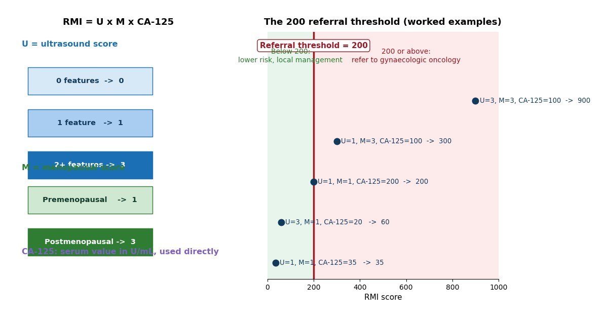
The formula is deliberately simple so it can be worked out at the bedside: RMI = U x M x CA-125, where CA-125 is the absolute serum value in U/mL. The two scores, U and M, are assigned as follows.
The ultrasound score U
Jacobs et al. identified five ultrasound features associated with malignancy, and each one present contributes one point: a multilocular cyst, solid areas within the mass, bilateral lesions (disease on both ovaries), ascites (free fluid in the abdomen), and intra-abdominal metastases. The total number of features present is then converted to the U score: no features gives U = 0, exactly one feature gives U = 1, and two or more features give U = 3. Because the three components are multiplied, a completely featureless scan (U = 0) always produces an RMI of 0, regardless of the CA-125 level. That is a deliberate property of the original index: without any suspicious ultrasound features, Jacobs found the risk of malignancy low enough that the other two factors should not move the decision.
The menopausal score M
Menopausal status is scored 1 for premenopausal women and 3 for postmenopausal women. Jacobs defined postmenopausal as 12 months or more of amenorrhoea, or an age of 50 years or more in a woman who has had a hysterectomy. The tripling of the score after the menopause reflects the biology: epithelial ovarian cancer is rare before the menopause and its incidence rises sharply afterwards, so the same ultrasound picture and the same CA-125 level mean something quite different in a 30 year old than in a 65 year old.
The CA-125 value
The serum CA-125 concentration in U/mL is used directly, without any transformation. The laboratory upper reference limit for CA-125 is commonly 35 U/mL, but the RMI does not use a raised-versus-normal dichotomy: the actual number goes into the multiplication. A postmenopausal woman with two ultrasound features (U = 3) and a CA-125 of 100 U/mL therefore has an RMI of 3 x 3 x 100 = 900, well above the referral threshold of 200. A premenopausal woman with one ultrasound feature (U = 1) and a CA-125 of 35 U/mL has an RMI of 1 x 1 x 35 = 35, comfortably below the threshold.
RMI-II, RMI-III and the other variants
The original Jacobs index is now usually called RMI-I, because three later variants have been proposed. Tingulstad and colleagues in Norway modified the scoring in 1996 to create RMI-II: the ultrasound score became 1 when zero or one features were present and 4 when two or more were present, while menopausal status became 1 or 4. The cut-off stayed at 200, and in their original 173 patients the sensitivity was 71% with a specificity of 96%. In 1999 the same group proposed RMI-III, which keeps the cut-off of 200 but scores U as 1 for zero or one feature and 3 for two or more, with M as 1 or 3.
The practical difference between the versions is what happens with a completely benign-looking scan. In RMI-I, U = 0 forces the entire product to zero, so a very high CA-125 cannot lift the score. In RMI-II and RMI-III the minimum U is 1, so the score always reflects the menopausal status and the CA-125 value even when the ultrasound shows nothing suspicious. Yamamoto and colleagues later proposed RMI-IV in 2009, which adds a fourth factor, tumour size (S), and uses a cut-off of 450, but it has been much less widely adopted. This calculator implements RMI-I, the original Jacobs formula, because it remains the most studied version and the one embedded in most guidelines and referral pathways.
How accurate is the RMI?
Validation studies of the RMI now number in the hundreds, with reported sensitivities between about 51% and 90% and specificities between about 51% and 97% at the 200 cut-off, reflecting different patient populations and study designs. The most informative estimates come from systematic reviews. Geomini and colleagues published a systematic review of risk scores for ovarian malignancy in Obstetrics and Gynecology in 2009. Kaijser and colleagues then pooled the individual studies in Human Reproduction Update in 2014: across 23 studies of the RMI at a cut-off of 200, the pooled sensitivity was 72% (95% confidence interval 67 to 76%) and the pooled specificity was 92% (89 to 93%). For RMI-II, across 15 studies, the pooled sensitivity was 75% (69 to 80%) and specificity 87% (84 to 90%). In plain terms, at the 200 threshold the index misses roughly a quarter of cancers but is correct in about nine out of ten benign cases.
Accuracy also depends on the type of tumour. The RMI performs best for epithelial ovarian cancers, which are the commonest malignant adnexal tumours and the ones that typically raise CA-125. It is notably less sensitive for borderline ovarian tumours and for non-epithelial cancers such as germ cell tumours, which may not raise CA-125 at all. The ultrasound component is also operator dependent: the same mass can be scored differently by a general sonographer and by an expert in gynaecological ultrasound, and studies using the IOTA (International Ovarian Tumor Analysis) criteria have shown expert pattern recognition outperforming the RMI. For these reasons the RMI is best understood as a triage tool for the general hospital setting, where it reliably separates the masses that clearly need the cancer centre from those that do not, rather than as a diagnostic instrument.
What the result means in practice
An RMI of 200 or more is the referral threshold used in the original study and in most subsequent practice: the woman should be referred to a gynaecologic oncology centre for assessment and, if surgery is needed, for the operation itself. This is not because an RMI of 200 means cancer is certain. It means the probability of malignancy is high enough that the potential benefit of specialist surgery outweighs the inconvenience of referral. In the original Jacobs study, patients above 200 had 42 times the background risk of cancer, a figure that has made the threshold durable for more than three decades.
An RMI below 200 indicates a lower risk, and such masses are usually managed in a general gynaecology unit with an appropriate operation for a presumed benign mass. A low score is reassuring but not a guarantee: a minority of cancers, particularly mucinous and non-epithelial tumours with normal CA-125, score below 200. Clinical judgement, the woman's symptoms, and the trajectory of the mass on repeat imaging all still matter. The RMI informs the referral decision; it never replaces the clinician.
The limits of CA-125 and the RMI
The weakest link in the RMI is the marker it multiplies by. CA-125 is raised in most epithelial ovarian cancers, but it is also raised in a long list of benign conditions: endometriosis, uterine fibroids, pelvic inflammatory disease, and even menstruation and early pregnancy. In a premenopausal woman with endometriosis, a CA-125 of several hundred U/mL is entirely possible with no malignancy present, which is one reason the menopausal score exists and why a raised CA-125 on its own should never be treated as a cancer diagnosis. Conversely, a normal CA-125 does not exclude cancer. Mucinous ovarian cancers often produce little CA-125, and non-epithelial tumours may produce none at all; such cancers can present with a low RMI.
The RMI also has defined boundaries of use. It was developed and validated in non-pregnant women with an adnexal mass detected clinically or on imaging, not as a screening test for asymptomatic women, and it should not be applied in pregnancy. It does not incorporate symptoms, family history, or newer markers such as HE4 (used in the ROMA algorithm), and it does not replace a careful clinical assessment. Treat the number as what it is: a well validated, deliberately simple aid for deciding where a woman with a pelvic mass should be treated.
References
- Jacobs I, Oram D, Fairbanks J, Turner J, Frost C, Grudzinskas JG. A risk of malignancy index incorporating CA 125, ultrasound and menopausal status for the accurate preoperative diagnosis of ovarian cancer. Br J Obstet Gynaecol. 1990;97(10):922-929. (Reported in Obstet Gynecol. 1990;75:1015-1022 in the original description cited by subsequent authors.)
- Tingulstad B, Hagen B, Skjeldestad FE, et al. Evaluation of a risk of malignancy index based on serum CA-125, ultrasound findings and menopausal status in the pre-operative diagnosis of pelvic masses. Br J Obstet Gynaecol. 1996;103(8):826-831. (RMI-II.)
- Tingulstad B, Hagen B, Skjeldestad FE, Halvorsen T, Nustad K, Onsrud M. The risk-of-malignancy index to evaluate potential ovarian cancers in local hospitals. Obstet Gynecol. 1999;93(3):448-452. (RMI-III.)
- Geomini P, Kruitwagen R, Bremer GL, Cnossen J, Mol BW. The accuracy of risk scores in predicting ovarian malignancy: a systematic review. Obstet Gynecol. 2009;113(2 Pt 1):384-394.
- Kaijser J, Sayasneh A, Van Hoorde K, et al. Presurgical diagnosis of adnexal tumours using mathematical models and scoring systems: a systematic review and meta-analysis. Hum Reprod Update. 2014;20(3):449-462.
- Yamamoto Y, Yamada R, Oguri H, et al. Comparison of four malignancy risk indices in the preoperative evaluation of patients with pelvic masses. Eur J Obstet Gynecol Reprod Biol. 2009;144(2):163-167. (RMI-IV.)
- American College of Obstetricians and Gynecologists
- WHO: Women's Health
Related calculators on Doctor With Data: all women's health calculators, the full calculator library.
Key takeaways
- The Risk of Malignancy Index (RMI) is a simple preoperative triage score for women with an adnexal (pelvic or ovarian) mass, published by Jacobs and colleagues in 1990.
- Give one point for each of five ultrasound features described by Jacobs et al.: a multilocular cyst, solid areas within the mass, bilateral lesions, ascites, and intra-abdominal metastases.
- Jacobs et al.
- An RMI of 200 or more is the accepted referral threshold: the mass should be assessed and operated on in a specialist gynaecologic oncology centre rather than a general hospital.
Frequently asked questions
What is the Risk of Malignancy Index?
The Risk of Malignancy Index (RMI) is a simple preoperative triage score for women with an adnexal (pelvic or ovarian) mass, published by Jacobs and colleagues in 1990. It multiplies three things: an ultrasound score U (0, 1 or 3), a menopausal status score M (1 for premenopausal, 3 for postmenopausal) and the serum CA-125 level in U/mL. A total of 200 or more flags a higher risk and signals referral to a gynaecologic oncology centre; a total below 200 is lower risk and can usually be managed locally. It is a referral aid, not a diagnosis.
How do I work out the ultrasound score U?
Give one point for each of five ultrasound features described by Jacobs et al.: a multilocular cyst, solid areas within the mass, bilateral lesions, ascites, and intra-abdominal metastases. If none of these features is present, U is 0. If exactly one is present, U is 1. If two or more are present, U is 3. Note that with U equal to 0 the RMI is always 0, whatever the CA-125 value, because the three components are multiplied.
What counts as postmenopausal for the RMI?
Jacobs et al. defined postmenopausal as 12 months or more of amenorrhoea, or an age of 50 years or more if the woman has had a hysterectomy. In that case M is 3; otherwise M is 1. This matters because ovarian cancer risk rises sharply after the menopause and the RMI weights it accordingly.
What does an RMI of 200 or more mean?
An RMI of 200 or more is the accepted referral threshold: the mass should be assessed and operated on in a specialist gynaecologic oncology centre rather than a general hospital. In the original study the cut-off had a sensitivity of 85.4% and a specificity of 96.9%, and across 23 studies in the Kaijser 2014 meta-analysis the pooled sensitivity was 72% and specificity 92%. It does not mean the mass is cancerous; it means the risk is high enough to need specialist care.
What is the difference between RMI-I, RMI-II and RMI-III?
RMI-I is the original Jacobs formula: U is 0, 1 or 3; M is 1 or 3; threshold 200. RMI-II, proposed by Tingulstad et al. in 1996, scores U as 1 for zero or one feature and 4 for two or more, and M as 1 or 4; threshold 200. RMI-III, from Tingulstad et al. in 1999, scores U as 1 for zero or one feature and 3 for two or more, and M as 1 or 3; threshold 200. The main practical difference is that the later versions never multiply by zero, so a completely benign-looking scan no longer forces the score to zero. This calculator implements RMI-I, the original and most widely studied version.
Can CA-125 be raised without cancer?
Yes, very commonly. CA-125 rises in benign gynaecological conditions such as endometriosis, uterine fibroids and pelvic inflammatory disease, and can also rise during menstruation and pregnancy. That is why a single CA-125 value is a weak test on its own and why the RMI combines it with ultrasound findings and menopausal status. Conversely, a normal CA-125 does not rule out malignancy, particularly for mucinous or non-epithelial ovarian tumours that may not raise the marker.
Medical disclaimer. This calculator is an educational tool only. It implements the published RMI-I formula for the preoperative triage of adnexal masses and does not provide medical advice, a diagnosis, or a treatment recommendation. A high or low score must be interpreted by a qualified clinician in the context of the full clinical picture. If you have a pelvic mass, persistent bloating, pelvic pain, or other concerning symptoms, see your doctor or gynaecologist promptly.