Why left bundle branch block hides a heart attack
The standard ECG diagnosis of acute myocardial infarction depends on recognising ST-segment elevation in the leads overlying the injured territory. That works when the ventricles depolarise normally. In left bundle branch block (LBBB), the left ventricle is activated late and abnormally, through slow cell-to-cell spread rather than the fast His-Purkinje system. The result is a wide, bizarre QRS complex and, crucially, secondary repolarisation abnormalities: the ST segments and T waves point in the opposite direction to the main QRS deflection.
This expected discordance is the camouflage. In a normal heart, ST elevation is a red flag; in LBBB, some degree of discordant ST elevation is simply part of the conduction pattern, so the ischaemic signal drowns in the background noise of the block itself. A clinician studying the tracing cannot easily tell whether a 3 mm ST elevation in a deeply negative complex is the usual secondary change of LBBB or the added primary change of an occluded artery. The same problem occurs in a ventricular-paced rhythm, where the paced beat produces a wide QRS with a similar discordant ST-T pattern.
What breaks the camouflage is concordance: an ST shift in the same direction as the QRS complex violates the expected pattern of LBBB and is never normal. That single insight is the physiological basis of the Sgarbossa criteria, published in 1996, which turned a gestalt impression into a weighted, testable score.
What the Sgarbossa criteria are
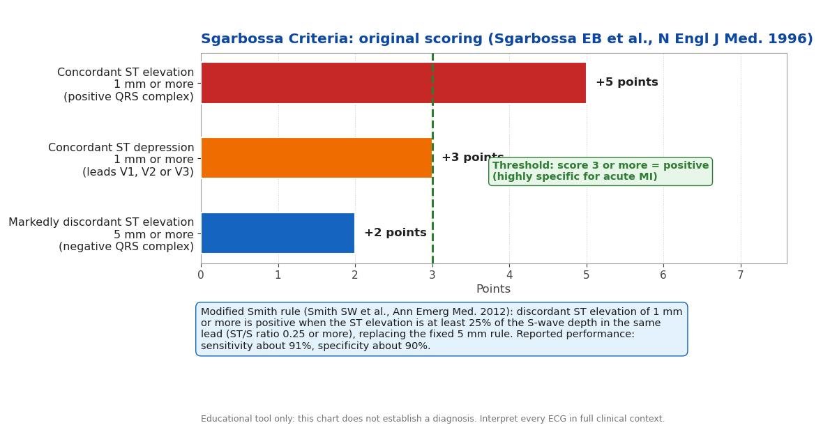
Elena Sgarbossa and colleagues derived the criteria from electrocardiograms collected during the GUSTO-1 trial, comparing 26 patients who had LBBB with enzyme-confirmed acute myocardial infarction against 141 patients who had LBBB without infarction (Sgarbossa EB et al., N Engl J Med. 1996;334:481-487). Using multivariate analysis, they identified three ECG features that independently predicted infarction and assigned each a weight reflecting its strength: 5 points, 3 points, and 2 points.
The points are added to give a total score out of 10, and a score of 3 or more is considered positive. The weighting encodes clinical reasoning: the most specific finding carries the most points, so a single striking abnormality can make the diagnosis, while weaker findings must combine. The criteria were designed for one specific clinical question, whether an adult with LBBB and symptoms suggesting acute coronary syndrome is having an MI, and they were never intended as a general screening test.
The three original criteria and their weights
- Concordant ST elevation of 1 mm or more in a lead with a positive QRS complex: 5 points. This is the strongest single criterion. In LBBB, a predominantly upright QRS should be followed by a discordant (depressed) ST segment, so elevation in the same direction as the QRS is profoundly abnormal and mirrors the classic ST-elevation pattern seen in a normally conducted heart. It is uncommon in uncomplicated LBBB, which is why it earns the full 5 points and is positive on its own.
- Concordant ST depression of 1 mm or more in lead V1, V2, or V3: 3 points. In LBBB, leads V1 to V3 normally show deep S waves with discordant, slightly elevated ST segments. ST depression there is concordant with the negative QRS and therefore abnormal; it is the mirror image of anterior ischaemia and is particularly associated with occlusion of the left anterior descending artery. Three points make it positive on its own as well.
- Markedly discordant ST elevation of 5 mm or more in a lead with a negative QRS complex: 2 points. Some discordant elevation is expected in LBBB, but 5 mm or more is excessive and suggests that an ischaemic injury current has been added to the secondary repolarisation change. Because modest discordant elevation can occur without infarction, this is the least specific finding and carries only 2 points, so it contributes to a positive score only in combination with another criterion.
All measurements are made in millimetres at the J point, where one small ECG square equals 1 mm. Note the asymmetry the authors built in: the two concordance criteria each reach the positive threshold alone, while the discordance criterion needs company.
The modified Sgarbossa rule: the Smith ST/S ratio
The weakest link in the original score is the fixed 5 mm cutoff for discordant elevation. Five millimetres is not equally excessive in every lead: in a lead with a very deep S wave, 5 mm of discordant elevation may be entirely proportional to the QRS voltage, while in a lead with a shallow S wave, even 3 mm can be disproportionate. Smith SW and colleagues addressed this in 2012 by replacing the absolute cutoff with a proportional one (Smith SW et al., Ann Emerg Med. 2012;60(6):766-776).
Under the modified rule, discordant ST elevation of at least 1 mm is positive when the ST elevation is at least 25 percent of the depth of the S wave in the same lead: an ST/S ratio of 0.25 or more. In the paper this is expressed as -0.25 or less, a sign convention meaning the ST segment deviates in the opposite direction to the S wave by at least a quarter of its depth. The other two criteria are unchanged, and the modified rule is positive if any one of the three is met.
In the original validation, the modified rule achieved a reported sensitivity of about 91% with a specificity of about 90%: a large gain in sensitivity over the original score of 3 or more, at a modest cost in specificity. This calculator computes both the original weighted score and the modified rule, and reports each verdict separately so you can see exactly which criterion drove the result.
How to use this calculator
Measure each ST deviation in millimetres at the J point, where one small square equals 1 mm, using the TP segment or the PR segment as the baseline as your local practice dictates, and be consistent across all leads. Enter the largest concordant ST elevation found in any lead with a predominantly positive QRS complex, or 0 if there is none. Then record whether concordant ST depression of 1 mm or more is present in V1, V2 or V3. Next, enter the largest discordant ST elevation in any lead with a predominantly negative QRS complex, or 0 if there is none; if you enter any value above 0 here, you must also enter the depth of the S wave in that same lead so the calculator can compute the ST/S ratio.
The calculator validates every entry before scoring: missing values and non-numeric entries produce named errors telling you exactly which field needs attention, and the S-wave depth is required whenever discordant ST elevation is entered. Press Calculate to see the original score out of 10 with its verdict, the modified Smith verdict with the computed ST/S ratio, and the list of criteria that contributed to each result.
Interpreting the result
An original Sgarbossa score of 3 or more is POSITIVE: in the derivation study this threshold was highly specific for acute myocardial infarction, with a reported specificity of approximately 90 to 98 percent depending on the threshold used. In a patient with ischaemic symptoms, a positive score should prompt the same urgent response as an ST-elevation pattern in a normally conducted heart: immediate cardiology involvement and activation of the reperfusion pathway.
A score below 3 is NEGATIVE by the original criteria, but a negative score does not rule out infarction. The original criteria have a reported sensitivity of only about 36 to 52 percent, so a substantial proportion of infarctions in LBBB score below 3. A negative result therefore means continue the workup, not stand down: serial ECGs and high-sensitivity troponins remain essential.
A POSITIVE modified Smith result carries a reported sensitivity of about 91% with a specificity of about 90%, making it a more sensitive but slightly less specific trigger than the original score. When the two rules disagree, the modified rule will more often be the positive one; that pattern is expected, and it is why the calculator names the criterion behind each verdict. Neither verdict is a diagnosis on its own: both must be interpreted alongside symptoms, haemodynamics, prior ECGs, and biomarker trends.
How accurate are the criteria?
The numbers matter because the stakes are reperfusion decisions. In the original 1996 study, a Sgarbossa score of 3 or more was highly specific for acute MI, with specificity reported at approximately 90 to 98 percent depending on the exact threshold applied, while sensitivity was approximately 36 to 52 percent (Sgarbossa EB et al., N Engl J Med. 1996;334:481-487). High specificity means a positive result is rarely wrong, which is exactly what you want before committing a patient to emergency reperfusion. Low sensitivity means a negative result is weak reassurance: many true infarctions will not reach 3 points.
The modified Smith rule shifted this balance, with a reported sensitivity of about 91% and specificity of about 90% (Smith SW et al., Ann Emerg Med. 2012;60(6):766-776). In practical terms, the modified rule misses far fewer infarctions but will also label more non-infarctions as positive. Both sets of figures come from selected study populations of patients with LBBB and suspected acute coronary syndrome, so they describe test performance in that setting, not in unselected ECGs. Apply the criteria to the patient in front of you only when the clinical question matches the one the studies asked: suspected acute MI in LBBB or a ventricular-paced rhythm.
Limitations
- Low sensitivity of the original score. A negative Sgarbossa score never excludes MI. The criteria must sit inside a pathway of serial ECGs and high-sensitivity troponin testing, and clinical suspicion should always overrule a negative score.
- Measurement precision. One millimetre is the difference between positive and negative for two of the three criteria, so baseline wander, artefact, and inconsistent J-point measurement can flip the result. Confirm the finding on a clean tracing before acting on it.
- LBBB is the validated setting. The criteria were derived and validated in left bundle branch block. The same QRS and ST logic is widely applied to ventricular-paced rhythms, which produce a similar wide-QRS discordant pattern, but the direct validation evidence there is thinner, so extra caution is warranted.
- Narrow intended population. The criteria answer a single question in adults with suspected acute coronary syndrome. They are not validated for screening asymptomatic people, for children, or for other conduction patterns such as right bundle branch block.
- Prior ECGs matter. A new concordant change is more alarming than a chronic one, and comparison with an old tracing is one of the most powerful diagnostic steps available. Always compare when a prior ECG exists.
- No score replaces clinical judgment. Haemodynamic instability, ongoing ischaemic pain, and cardiology input drive reperfusion decisions. The Sgarbossa criteria are an aid to ECG interpretation, not a substitute for it.