Why suspected DVT starts with a probability, not a scan
A painful, swollen leg is one of the most common reasons people present to emergency departments and urgent care, and only a minority of those legs contain a clot. Cellulitis, a ruptured Baker cyst, superficial thrombophlebitis, a torn calf muscle, and chronic venous insufficiency can all look remarkably like deep vein thrombosis. Compression ultrasound is the definitive test, but scanning every swollen leg would swamp imaging departments, delay genuinely urgent cases, and still leave clinicians unsure what to do with borderline findings. The smarter strategy is to estimate the probability of DVT before ordering anything, because that probability changes what every subsequent test means.
This is where a clinical prediction rule earns its place. A D-dimer blood test is very sensitive but poorly specific: a positive result raises many possibilities, while a negative result is only reassuring when the pretest probability was already low. The Wells score supplies that pretest probability in a structured, reproducible way. A low score plus a negative D-dimer rules out DVT safely without any imaging, while a high score sends the patient straight to ultrasound. The score therefore does not diagnose anything on its own; it routes each patient to the correct next step and spares low-risk patients an unnecessary scan.
What the Wells score is
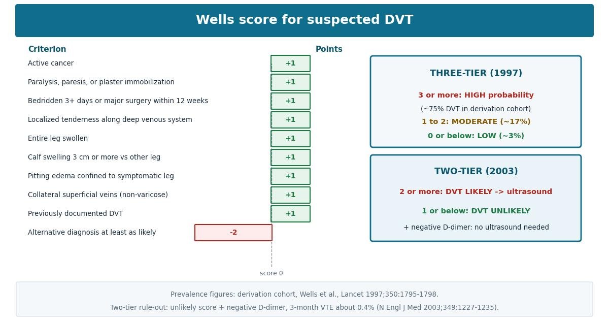
The score was developed by Philip Wells and colleagues in Ottawa and published in 1997 (Wells PS, Anderson DR, Bormanis J, et al. Value of assessment of pretest probability of deep-vein thrombosis in clinical management. Lancet. 1997;350(9094):1795-1798). The team studied outpatients referred with suspected DVT, identified the clinical features that independently predicted a confirmed clot, and combined them into a simple additive score. Nine features each add one point, and a single clinical judgment, that an alternative diagnosis is at least as likely as DVT, subtracts two points, giving a total that ranges from -2 to 9.
The original model divided patients into three probability bands: high, moderate, and low. In 2003 the same group simplified the bands into two, DVT likely and DVT unlikely, to support a clean management rule for use with D-dimer testing (Wells PS, Anderson DR, Rodger M, et al. Evaluation of D-dimer in the diagnosis of suspected deep-vein thrombosis. N Engl J Med. 2003;349:1227-1235). The items and weights never changed, only the cutoffs. Both versions remain in widespread clinical use, and this calculator reports both interpretations from the same ten answers.
The nine one-point criteria
- Active cancer: +1 point. Cancer counts if the patient is receiving treatment or has received treatment within the last six months, or is receiving palliative care. Malignancy is one of the strongest risk factors for venous thromboembolism because tumours and their treatment promote clotting through several mechanisms, so this item flags a fundamentally higher-risk patient.
- Paralysis, paresis, or recent plaster immobilization of the lower extremities: +1 point. Anything that stops the calf muscle pump from squeezing blood back toward the heart lets blood stagnate in the deep veins of the leg. This includes a leg in plaster, a paralysed or paretic limb, and any other recent immobilization of the lower extremity.
- Recently bedridden for 3 days or more, or major surgery within the last 12 weeks: +1 point. Prolonged bed rest and the aftermath of major surgery combine venous stasis with the prothrombotic effects of tissue injury. The original definition of major surgery is surgery performed under general or regional anaesthesia within the preceding twelve weeks.
- Localized tenderness along the distribution of the deep venous system: +1 point. Tenderness that follows the course of the deep veins, typically along the calf or the medial thigh, rather than diffuse or superficial tenderness. This is a bedside finding that requires the examiner to palpate deliberately along the venous anatomy.
- Entire leg swollen: +1 point. Swelling that involves the whole leg suggests obstruction of a large proximal vein, which is the pattern most characteristic of an extensive DVT. This is distinct from swelling confined to the calf, which is captured by the next item.
- Calf swelling 3 cm or more compared with the asymptomatic leg: +1 point. Measure the calf circumference 10 cm below the tibial tuberosity on both legs; a difference of 3 cm or greater scores the point. The fixed measurement site and the 3 cm threshold keep this objective rather than a matter of impression.
- Pitting edema confined to the symptomatic leg: +1 point. Pitting edema that is present only in the symptomatic leg, not bilateral. Bilateral edema points toward systemic causes such as heart failure or hypoalbuminaemia, whereas strictly unilateral edema is the pattern expected with a venous obstruction.
- Collateral superficial veins, non-varicose: +1 point. Visible distended superficial veins that are not ordinary varicose veins. They represent blood finding alternative routes around a blocked deep system, and their presence implies the obstruction has been there long enough for collaterals to develop.
- Previously documented DVT: +1 point. A prior confirmed episode of deep vein thrombosis. Previous clot damages venous valves and leaves residual thrombus, both of which predispose to recurrence, so a history of DVT independently raises the probability that the current presentation is another one.
The minus-two point criterion
The tenth item is unlike the other nine: it is a judgment, not a finding. If an alternative diagnosis is at least as likely as DVT, subtract two points. Typical alternatives include cellulitis, a ruptured Baker cyst, superficial thrombophlebitis, a calf haematoma after minor trauma, and chronic venous disease. This item carries the largest single weight in the score because, in the original derivation, it was the strongest predictor that the patient did not have DVT. It is also the most subjective item, which is why the instructions demand honesty: the alternative must genuinely be at least as likely, not merely possible. A casual minus two applied to every ambiguous case will systematically underestimate risk.
How to use this calculator
Work through the ten questions in order, answering Yes or No to each. The calculator requires an answer to every question: if you leave any unanswered, it will name the missing item and refuse to score until you complete it, because a half-answered Wells score is worse than no score at all. Measure the calf circumference as described rather than guessing, and decide the alternative-diagnosis question only after a proper examination, not before.
Press Calculate to see the total score from -2 to 9, the list of items that contributed points, and both interpretations: the three-tier model (high, moderate, or low probability) and the two-tier model (DVT likely or DVT unlikely). Use the result to choose the next step: high or likely scores need compression ultrasound promptly, while low or unlikely scores open the D-dimer rule-out pathway described below.
Interpreting the result: the three-tier model
The original 1997 model defines three bands. A score of 3 or more is high probability: in the derivation cohort of outpatients with suspected DVT, about 75 percent of high-probability patients had DVT confirmed. A score of 1 or 2 is moderate probability, with a confirmed DVT prevalence of about 17 percent in the derivation study. A score of 0 or below is low probability, with a prevalence of about 3 percent.
These percentages describe the derivation population, not a universal law, but they explain the management logic. A high-probability patient should proceed directly to compression ultrasound, because even a negative D-dimer would not be reassuring enough at that pretest probability. A low-probability patient is the ideal candidate for the D-dimer rule-out pathway: if the D-dimer is negative, DVT is excluded without imaging. Moderate-probability patients sit in between and are managed according to local protocols, usually with D-dimer testing followed by ultrasound if the D-dimer is positive.
Interpreting the result: the two-tier model
The simplified 2003 model has only two bands: a score of 2 or more means DVT likely, and a score of 1 or below means DVT unlikely. Note how the cutoffs shift relative to the three-tier model: a score of exactly 2 is moderate in the three-tier system but likely in the two-tier system, which is intentional, because the two-tier model is designed to be paired with D-dimer in a single management rule.
The rule, validated in the 2003 Wells management study, is straightforward: in patients with an unlikely score, a negative highly sensitive D-dimer excludes DVT and ultrasound can be safely withheld. In that study, the 3-month rate of venous thromboembolism among unlikely-score patients with a negative D-dimer was about 0.4 percent, a miss rate low enough to justify skipping the scan. Everyone else, meaning likely scores regardless of D-dimer, and unlikely scores with a positive D-dimer, proceeds to compression ultrasound. The two-tier model trades some granularity for decisiveness, which is why many emergency departments prefer it.
What happens after the score: D-dimer and compression ultrasound
The Wells score is the first half of a diagnostic pathway; D-dimer and ultrasound are the second half. D-dimer is a fibrin breakdown product that rises whenever clot is forming and breaking down anywhere in the body. A highly sensitive D-dimer assay misses very few DVTs, which makes a negative result powerful, but it is elevated by cancer, infection, inflammation, recent surgery, pregnancy, and advancing age, so a positive result is common and non-specific. That asymmetry is exactly why the test only rules out DVT when the pretest probability is already low or unlikely.
Compression ultrasound of the proximal leg veins is the standard confirmatory test: the sonographer presses the probe on the vein, and a normal vein collapses completely while a vein containing thrombus does not. When the first ultrasound is negative but suspicion remains high, guidelines commonly advise repeating the proximal scan after about a week, because a small calf thrombus can extend proximally over days. Whole-leg ultrasound, which also examines the calf veins, is an alternative where available. Whichever pathway is used, patients sent home without a diagnosis need clear safety-netting advice about what should bring them back.
How accurate is the Wells score?
The most quoted performance figures are the derivation prevalences: about 75 percent DVT in the high-probability band, 17 percent in the moderate band, and 3 percent in the low band (Wells PS et al., Lancet. 1997;350(9094):1795-1798). These numbers describe how well the score stratified risk in the population it was built from, which was outpatients referred with suspected DVT. The score has since been validated in many independent cohorts, and while the exact percentages vary between settings, the ordering is consistent: the high band always contains far more DVT than the low band.
The strongest management evidence comes from the 2003 study that paired the two-tier score with D-dimer: withholding ultrasound in unlikely-score patients with a negative D-dimer produced a 3-month venous thromboembolism rate of about 0.4 percent (Wells PS et al., N Engl J Med. 2003;349:1227-1235). That is the figure that justifies the rule-out pathway used in emergency departments worldwide. Accuracy in practice depends heavily on honest application of the alternative-diagnosis item: applied carelessly, the minus two points deflate scores and push patients who need imaging into the rule-out pathway.
Limitations
- Built for suspected first proximal DVT of the leg in outpatients. The derivation and most validation cohorts were outpatients with a suspected first episode. Performance is less certain in hospital inpatients, in recurrent DVT where old residual thrombus confuses ultrasound, and in special populations.
- Not for pregnancy or the upper limb. D-dimer is physiologically elevated in pregnancy and the score was not derived there, so suspected DVT in pregnancy follows different guidance with specialist input. Suspected upper-extremity DVT needs its own prediction tools, such as the Constans score, not the Wells leg score.
- The minus-two item is subjective. Clinical judgment varies between examiners, and this single item can move a patient across two bands. If two clinicians would disagree about the alternative diagnosis, the safer reading is usually the one that does not subtract the points.
- D-dimer is easily positive. Age, cancer, infection, inflammation, and recent surgery all raise D-dimer, so a positive result in a low-risk patient usually means proceed to ultrasound, not start treatment. Age-adjusted D-dimer cutoffs exist in some protocols to reduce false positives in older patients.
- A low score never excludes DVT on its own. The rule-out requires the low or unlikely score together with a negative highly sensitive D-dimer. A score of 0 with a positive D-dimer still needs imaging, and a high score needs imaging regardless of the D-dimer result.
- No score replaces clinical judgment. If the presentation suggests pulmonary embolism rather than isolated leg DVT, use a PE prediction rule instead, and unstable or deteriorating patients need urgent senior assessment whatever the score says.