Wells Score Calculator for Suspected Pulmonary Embolism
In short: Wells score calculator for suspected pulmonary embolism: the 7-variable clinical prediction rule with both the three-tier (low, intermediate, high) and two-tier (PE unlikely, PE likely) interpretations. Use the calculator above, then read the guide below to interpret your result and its limitations.
Estimate the pretest probability of pulmonary embolism with the seven-variable Wells clinical prediction rule. The calculator reports both the three-tier interpretation (low, intermediate, high probability) and the two-tier interpretation (PE unlikely, PE likely).
Calculator
What the Wells score is
The Wells score is a clinical prediction rule that estimates the pretest probability of pulmonary embolism in patients with suspected disease. It was derived by Wells PS and colleagues and published in the Annals of Internal Medicine in 1998 (Wells PS et al., Ann Intern Med. 1998;129(12):997-1005; doi:10.7326/0003-4819-129-12-199812150-00002; PMID 9867786), then validated in an independent emergency department cohort published in the same journal in 2001 (Wells PS et al., Ann Intern Med. 2001;135(2):98-107; doi:10.7326/0003-4819-135-2-200107170-00010; PMID 11453709).
Pretest probability matters because no test result can be interpreted on its own. A D-dimer, for example, has a high negative predictive value only when the starting probability of disease is already low: the same negative D-dimer is reassuring in a low-probability patient and unsafe in a high-probability one. The Wells score formalises the bedside assessment so that the result of D-dimer testing or imaging is combined with a measured starting probability rather than a gut feeling. That is why every major diagnostic pathway for suspected pulmonary embolism, including the ones validated in the Christopher study (van Belle A et al., JAMA. 2006;295(2):172-179; PMID 16403929), begins with a Wells score or an equivalent clinical probability assessment.
Pulmonary embolism is worth this effort because it is common and frequently fatal when missed. Venous thromboembolism is estimated to affect around 900,000 people in the United States each year, with 60,000 to 100,000 deaths attributed to it (Beckman MG et al., Am J Prev Med. 2010;38(4 Suppl):S495-S501; PMID 20331949). A validated rule that tells the clinician which patients can be safely discharged after a D-dimer and which need a CT pulmonary angiogram reduces both missed diagnoses and unnecessary radiation and contrast exposure.
The seven criteria and their weights
The score is the sum of points from seven clinical variables. The weights reflect how strongly each variable predicted pulmonary embolism in the derivation study:
- Clinical signs and symptoms of deep vein thrombosis (3 points). This requires a minimum of leg swelling and pain on palpation of the deep veins. It is the single strongest bedside clue because pulmonary embolism and deep vein thrombosis are two presentations of the same disease, venous thromboembolism: most pulmonary emboli originate as thrombus in the deep veins of the legs.
- Pulmonary embolism is the number one diagnosis, or equally likely (3 points). No alternative diagnosis is more likely than pulmonary embolism. This is the most subjective item in the rule and depends on the clinician's overall assessment after history, examination, chest radiograph, and electrocardiogram. It carries the same weight as objective DVT signs, which is one reason the rule's reproducibility between clinicians is imperfect.
- Heart rate above 100 beats per minute (1.5 points). Tachycardia is a common physiological response to pulmonary embolism, reflecting the right ventricle working against a suddenly increased pulmonary vascular resistance.
- Immobilisation of 3 days or more, or surgery in the previous 4 weeks (1.5 points). Venous stasis is one of the three components of Virchow's triad, and prolonged immobility or recent surgery markedly increases thrombotic risk. Either condition alone scores the points; the original wording is immobilisation of at least 3 days or surgery in the previous 4 weeks.
- Previously objectively confirmed deep vein thrombosis or pulmonary embolism (1.5 points). A prior venous thromboembolic event is a strong risk factor for recurrence. The confirmation must have been objective, meaning imaging or another definitive test, not a clinical impression.
- Haemoptysis (1 point). Coughing up blood occurs when an embolus causes pulmonary infarction with alveolar haemorrhage. It is uncommon but, when present in a patient with pleuritic chest pain, should raise suspicion.
- Malignancy (1 point). Defined as cancer on treatment, treated in the previous 6 months, or palliative care. Malignancy creates a hypercoagulable state, the second component of Virchow's triad, and is a well-established risk factor for venous thromboembolism.
The maximum possible score is 12.5 points. Scores advance in half-point steps, so exact thresholds matter: a score of exactly 4 is PE unlikely, while 4.5 is PE likely.
How to interpret the result
This calculator reports both published interpretation schemes. Use the one your local diagnostic pathway specifies; they are two views of the same score.
| Score | Three-tier interpretation | PE prevalence in the derivation group |
|---|---|---|
| Below 2 | Low probability | 3.6% |
| 2 to 6 | Intermediate probability | 20.5% |
| Above 6 | High probability | 66.7% |
| Score | Two-tier interpretation | PE prevalence in the derivation group |
|---|---|---|
| 4 or below | PE unlikely | 5.1% |
| Above 4 | PE likely | 39.1% |
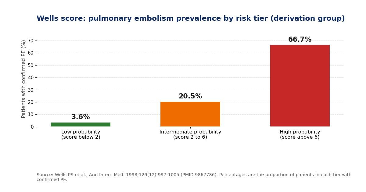
The prevalence figures come from the derivation study (Wells PS et al., Ann Intern Med. 1998;129(12):997-1005; PMID 9867786). They describe the proportion of patients in each group who turned out to have pulmonary embolism, which is why the score is called a pretest probability estimate. A high-probability score is not a diagnosis: roughly one in three patients in the high tier did not have pulmonary embolism. Equally, a low score does not exclude disease: about 1 in 28 low-probability patients in the derivation study had it, which is why the score must be combined with D-dimer testing rather than used alone.
What to do with the result
The Wells score guides the next test, it does not replace it. The standard two-tier pathway, validated in the Christopher study of 3,306 patients with suspected pulmonary embolism (van Belle A et al., JAMA. 2006;295(2):172-179; PMID 16403929), works as follows:
- PE unlikely (4 or below) plus a negative D-dimer: pulmonary embolism is considered excluded, and no imaging is needed. In the Christopher study this pathway was safe, with a very low rate of venous thromboembolism during 3 months of follow-up.
- PE unlikely with a positive D-dimer: proceed to CT pulmonary angiography.
- PE likely (above 4): proceed directly to CT pulmonary angiography. A D-dimer is not used to exclude disease in this group, because a negative result leaves the post-test probability too high.
The D-dimer cutoff used with this pathway is usually 500 micrograms per litre fibrinogen-equivalent units with a sensitive assay. The ADJUST-PE study (Righini M et al., JAMA. 2014;311(11):1117-1124; PMID 24577510) showed that an age-adjusted cutoff, defined as the patient's age multiplied by 10 micrograms per litre for patients over 50, increased the number of patients in whom pulmonary embolism could be excluded without imaging, without increasing missed events. The YEARS study (van der Hulle CA et al., Lancet. 2017;390(10091):289-297; PMID 28549662) combined a simplified Wells score, in which each of the seven items counts as 1 point, with variable D-dimer thresholds and safely reduced the number of CT scans: among 3,465 patients managed this way, CT pulmonary angiography was avoided in about 48%, and the 3-month rate of symptomatic venous thromboembolism was 0.61% (95% confidence interval 0.36 to 0.96).
Patients who are haemodynamically unstable with suspected massive pulmonary embolism should not wait for a score: they need immediate resuscitation and senior input, with bedside echocardiography and CT as the clinical situation allows.
How accurate is the Wells score?
The derivation study classified patients into low, intermediate, and high probability groups with confirmed pulmonary embolism rates of 3.6%, 20.5%, and 66.7% respectively (Wells PS et al., 1998; PMID 9867786). The 2001 validation study confirmed that the rule performed similarly in a new emergency department population and that the two-tier version combined with D-dimer testing could safely exclude pulmonary embolism (Wells PS et al., 2001; PMID 11453709). A later individual-patient-data meta-analysis of the Wells rule combined with D-dimer testing (van Es N et al., Ann Intern Med. 2016;165(4):253-264; PMID 27271328) confirmed that the pathway performs consistently across settings, supporting its place in guidelines worldwide.
The rule's main weakness is the subjective item: whether pulmonary embolism is the most likely diagnosis depends on the clinician, and agreement between clinicians on this item is only moderate. This is a feature as much as a flaw, since it lets experienced judgment contribute, but it means two clinicians can legitimately score the same patient differently.
Limitations: when not to use the Wells score
- Pregnancy and the postpartum period. The rule was not derived or validated in pregnant or postpartum patients. A prospective study concluded that neither the Wells score nor the revised Geneva score could safely rule out pulmonary embolism in that population (Touhami O et al., Eur J Obstet Gynecol Reprod Biol. 2018;221:166-171). Suspected pulmonary embolism in pregnancy needs a dedicated diagnostic pathway.
- Patients already on therapeutic anticoagulation. The score and its D-dimer pathway do not apply when the patient is anticoagulated, because the pretest probability and test characteristics change.
- Inpatients and the critically ill. The rule was derived largely in emergency department and outpatient populations. Hospitalised patients have a higher baseline prevalence of thromboembolism and different competing diagnoses, so the score should be applied with extra caution.
- It does not replace clinical judgment. A low score in a patient whose story strongly suggests pulmonary embolism should not override the clinician's concern, and a high score does not confirm the diagnosis. The score is one input to a diagnostic strategy, never the whole strategy.
Worked examples
Example 1. A 62-year-old man presents with sudden pleuritic chest pain and shortness of breath. He has a swollen, tender left calf (3 points), no more likely alternative diagnosis (3 points), and a heart rate of 112 (1.5 points). Total: 7.5, which is high probability in the three-tier scheme and PE likely in the two-tier scheme. He should proceed directly to CT pulmonary angiography.
Example 2. A 34-year-old woman has pleuritic chest pain after a long flight. She had foot surgery 3 weeks ago (1.5 points) and a heart rate of 104 (1.5 points), with no DVT signs, no prior VTE, no haemoptysis, and no malignancy. Total: 3.0, which is intermediate probability and PE unlikely. The correct next step is D-dimer testing: a negative result excludes pulmonary embolism, a positive result leads to imaging.
Example 3. A 45-year-old man has mild, atypical chest discomfort. He had a deep vein thrombosis confirmed by ultrasound 2 years ago (1.5 points) and reports a small amount of haemoptysis this morning (1 point), with a heart rate of 96. Total: 2.5, intermediate probability and PE unlikely. He needs a D-dimer and, if positive, imaging.
Wells score versus the revised Geneva score
The main alternative is the revised Geneva score (Le Gal G et al., Ann Intern Med. 2006;144(3):165-171; PMID 16461960), which uses fully objective variables including age, heart rate bands, and unilateral leg findings, with no subjective judgment item. The revised Geneva score is therefore more reproducible between clinicians, while the Wells score lets the clinician's overall impression contribute through the PE-is-most-likely item. Both are endorsed by guidelines, both are combined with D-dimer testing in the same way, and individual-patient-data comparisons show broadly similar performance. The choice between them is usually a matter of local protocol rather than evidence that one is clearly superior.
References
- Wells PS, Ginsberg JS, Anderson DR, Kearon C, Gent M, Turpie AG, Bormanis J, Weitz J, Chamberlain M, Bowie D, Barnes D, Hirsh J. Use of a clinical model for safe management of patients with suspected pulmonary embolism. Ann Intern Med. 1998;129(12):997-1005. doi: 10.7326/0003-4819-129-12-199812150-00002. PMID: 9867786.
- Wells PS, Anderson DR, Rodger M, Stiell I, Dreyer JF, Barnes D, Forgie M, Kovacs G, Ward J, Kovacs MJ. Excluding pulmonary embolism at the bedside without diagnostic imaging: management of patients with suspected pulmonary embolism presenting to the emergency department by using a simple clinical model and d-dimer. Ann Intern Med. 2001;135(2):98-107. doi: 10.7326/0003-4819-135-2-200107170-00010. PMID: 11453709.
- van Belle A, Buller HR, Huisman MV, Huisman PM, Kaasjager K, Kamphuisen PW, Kramer MH, Kruip MJ, Kwakkel-van Erp JM, Leebeek FW, Nijkeuter M, Prins MH, Sohne M, Tick LW; Christopher Study Investigators. Effectiveness of managing suspected pulmonary embolism using an algorithm combining clinical probability, D-dimer testing, and computed tomography. JAMA. 2006;295(2):172-179. doi: 10.1001/jama.295.2.172. PMID: 16403929.
- Righini M, Van Es J, Den Exter PL, Roy PM, Verschuren F, Ghuysen A, Rutschmann OT, Sanchez O, Jaffrelot M, Trinh-Duc A, Le Gall C, Moustafa F, Principe A, Van Houten AA, Ten Wolde M, Douma RA, Hazelaar G, Erkens PM, Van Kralingen KW, Grootenboers MJ, Durian MF, Cheung YW, Meyer G, Bounameaux H, Huisman MV, Kamphuisen PW, Le Gal G. Age-adjusted D-dimer cutoff levels to rule out pulmonary embolism: the ADJUST-PE study. JAMA. 2014;311(11):1117-1124. doi: 10.1001/jama.2014.2135. PMID: 24577510.
- van der Hulle CA, Dronkers CEA, Huisman MV, et al. Simplified diagnostic management of suspected pulmonary embolism (the YEARS study): a prospective, multicentre, cohort study. Lancet. 2017;390(10091):289-297. doi: 10.1016/S0140-6736(17)30885-1. PMID: 28549662.
- van Es N, van der Hulle CA, van Es J, den Exter PL, Douma RA, Goekoop RJ, Mos IC, Galipienzo J, Kamphuisen PW, Huisman MV, Klok FA, Buller HR, Bossuyt PM. Wells rule and D-dimer testing to rule out pulmonary embolism: a systematic review and individual-patient data meta-analysis. Ann Intern Med. 2016;165(4):253-264. doi: 10.7326/M16-0031. PMID: 27271328.
- Le Gal G, Righini M, Roy PM, Sanchez O, Aujesky D, Bounameaux H, Perrier A. Prediction of pulmonary embolism in the emergency department: the revised Geneva score. Ann Intern Med. 2006;144(3):165-171. doi: 10.7326/0003-4819-144-3-200602070-00007. PMID: 16461960.
- Beckman MG, Hooper WC, Critchley SE, Ortel TL. Venous thromboembolism: a public health concern. Am J Prev Med. 2010;38(4 Suppl):S495-S501. doi: 10.1016/j.amepre.2009.12.017. PMID: 20331949.
- American College of Emergency Physicians
- NICE Guidance
Key takeaways
- A score of 5 falls in the intermediate probability tier (2 to 6) and, under the two-tier scheme, in the PE likely group (above 4).
- No.
- Both use the same seven weighted items and the same total.
- No.
Frequently asked questions
What does a Wells score of 5 mean for suspected pulmonary embolism?
A score of 5 falls in the intermediate probability tier (2 to 6) and, under the two-tier scheme, in the PE likely group (above 4). In the original derivation group, about 20.5% of patients with a score of 2 to 6 had confirmed pulmonary embolism. It is a pretest probability estimate, not a diagnosis: a score of 5 means imaging, usually CT pulmonary angiography, is needed, and the treating clinician should not rely on a D-dimer alone in this range.
Can the Wells score be used in a patient already taking anticoagulants?
No. The Wells rule was derived and validated in patients with suspected first or recurrent pulmonary embolism who were not on therapeutic anticoagulation at the time of assessment. Anticoagulation changes the pretest probability and the behaviour of D-dimer testing, so the score and its cutoffs do not apply in that setting. A patient on therapeutic anticoagulation with new suspected pulmonary embolism needs direct clinical assessment and imaging as the team decides, not a Wells score.
What is the difference between the three-tier and two-tier Wells score?
Both use the same seven weighted items and the same total. The three-tier scheme groups scores into low (below 2), intermediate (2 to 6), and high (above 6) clinical probability. The two-tier scheme collapses this into PE unlikely (4 or below) and PE likely (above 4). The two-tier version was developed so that a PE unlikely result combined with a negative D-dimer could safely exclude pulmonary embolism without imaging, which is the pathway validated in the Christopher study and now used in most emergency departments.
Can a negative D-dimer rule out pulmonary embolism in a high-probability patient?
No. D-dimer testing is only validated as a rule-out test in patients with low or PE-unlikely pretest probability. In high-probability patients the post-test probability after a negative D-dimer remains too high to be safe, so imaging is required regardless of the D-dimer result. The safe exclusion pathway is: Wells PE unlikely (4 or below) plus a negative D-dimer by a sensitive assay; anything else needs imaging or specialist input.
Is the Wells score validated for use in pregnancy?
The Wells score was not derived or validated in pregnant or postpartum patients, and the evidence suggests it does not perform reliably there. A prospective study in pregnant and postpartum women concluded that neither the Wells score nor the revised Geneva score could safely rule out pulmonary embolism in that population (Touhami O et al., Eur J Obstet Gynecol Reprod Biol. 2018;221:166-171). Suspected pulmonary embolism in pregnancy should follow a dedicated diagnostic pathway with obstetric and radiology input, not a standard Wells score.
Does the Wells score diagnose pulmonary embolism?
No. The Wells score estimates the pretest probability of pulmonary embolism before definitive testing; it is a decision aid, not a diagnostic test. Even a high score does not confirm pulmonary embolism, since about one in three patients in the high-probability tier of the derivation study did not have it, and a low score does not guarantee its absence. Diagnosis rests on imaging, usually CT pulmonary angiography, interpreted alongside the clinical picture, D-dimer, and the judgment of the treating team.
Medical disclaimer
This calculator is an educational decision aid for clinicians and students. It estimates pretest probability and does not diagnose pulmonary embolism, rule it out, or replace clinical judgment. Suspected pulmonary embolism is a medical emergency: management decisions must be made by a qualified clinician using the full clinical picture, appropriate D-dimer assays, and imaging. If you have symptoms such as sudden breathlessness, chest pain, or coughing up blood, seek urgent medical care.