What is the revised Geneva score?
Pulmonary embolism is a common and potentially fatal condition in which a blood clot, usually from a deep vein thrombosis in the leg, travels to the lungs. Its symptoms, chest pain, breathlessness, a fast heart rate, and coughing up blood, overlap with many other conditions, so clinicians cannot diagnose it reliably from symptoms alone. Definitive imaging, usually CT pulmonary angiography, exposes patients to radiation and contrast dye, so doctors first estimate the clinical (pretest) probability that PE is present and then choose the next test accordingly. The revised Geneva score is one of the two standard tools for that estimate, alongside the Wells score.
The score was constructed by Le Gal and colleagues and published in the Annals of Internal Medicine in 2006. The team started from the original Geneva score of Wicki and colleagues, published in 2001, which performed well but required arterial blood gas results, a test that is painful, slow, and often unavailable at the bedside. The stated objective of the new work was to construct a simple score based entirely on clinical variables and independent of physicians' implicit judgment. The authors derived the score from consecutive emergency department patients with clinically suspected pulmonary embolism at three university hospitals in Europe, assigning points to each variable according to its regression coefficient, and then validated it in an independent cohort of similar patients.
That origin explains the score's defining feature. Every item is something the clinician can observe or ask about at the bedside: age, past history, current symptoms, and two examination findings, including the heart rate. There is no laboratory value, no imaging result, and no room for the clinician's overall impression. The score is fully standardised, which means two clinicians assessing the same patient should arrive at the same total. This is the deliberate contrast with the Wells score, whose best-known item awards points when the clinician judges that pulmonary embolism is the most likely diagnosis. That judgment, sometimes called gestalt, is accurate in experienced hands but cannot be standardised, taught, or audited in the same way. The revised Geneva score was built to remove it, and subsequent comparisons have shown it performs similarly to the Wells score without any subjective component.
The eight variables and their points
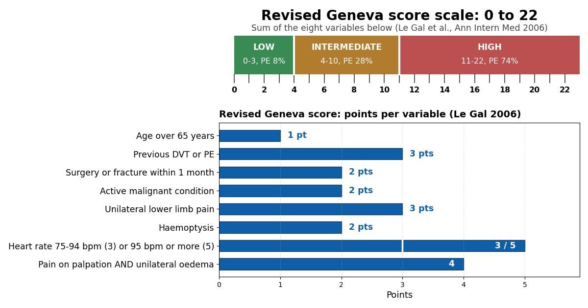
The score has eight variables. Their points are added, so the total ranges from 0 to 22. The variables and their points, exactly as published by Le Gal and colleagues, are:
- Age over 65 years: 1 point. The risk of venous thromboembolism rises with age, so older patients start with a point before any symptoms are considered. Note the strict cut point: a patient aged exactly 65 does not receive this point.
- Previous deep vein thrombosis or pulmonary embolism: 3 points. A prior clot is one of the strongest predictors of a new one, which is why this item carries the joint-highest weight among the history items.
- Surgery or fracture within the past month: 2 points. Recent surgery (particularly under general anaesthesia) or a fracture, especially of the lower limb, promotes clot formation through vessel injury and immobilisation.
- Active malignant condition: 2 points. Cancer creates a prothrombotic state, and active malignancy is a well-established risk factor for venous thromboembolism.
- Unilateral lower limb pain: 3 points. Pain in one leg suggests a deep vein thrombosis as the source of a possible embolus, and it scores as heavily as a previous clot.
- Haemoptysis: 2 points. Coughing up blood reflects lung infarction or congestion downstream of an embolus and is a classic, though uncommon, presenting symptom.
- Heart rate 75 to 94 beats per minute: 3 points; 95 beats per minute or more: 5 points. Tachycardia is the only variable with two positive levels, and the 5-point top level makes it the single highest-weighted item in the score. A heart rate below 75 scores nothing.
- Pain on lower-limb deep venous palpation combined with unilateral oedema: 4 points. This is the only compound item: the 4 points are awarded only when both findings are present. A patient with palpation pain but no swelling, or swelling without palpation pain, scores nothing for this item. This both-required rule is a common source of scoring mistakes, so it deserves deliberate checking.
Two features of the weighting are worth noticing. First, the history items (previous clot, recent surgery or fracture, active cancer) capture the patient's baseline thrombotic risk, while the symptom and sign items (leg pain, haemoptysis, heart rate, palpation pain with oedema) capture the current presentation. Second, the heaviest items, previous venous thromboembolism, unilateral leg pain, marked tachycardia, and the combined leg examination finding, are exactly the features an experienced clinician would weigh most heavily, which is part of why the score feels clinically sensible.
Adding up the score: the three probability bands
The points are summed to give a total between 0 and 22. A total of 0 to 3 means low pretest probability of PE. A total of 4 to 10 means intermediate pretest probability. A total of 11 or more means high pretest probability. These cut points come from the original Le Gal study and are used unchanged in current practice.
In the validation cohort, pulmonary embolism was ultimately confirmed in 8% of low-probability patients, 28% of intermediate-probability patients, and 74% of high-probability patients. These percentages describe how often PE was confirmed in each band during validation; they are not a personalised prediction for an individual patient, but they show why the bands matter. The separation between them is wide: the low band is low enough that a negative blood test can safely close the workup, while the high band is high enough that even a negative blood test cannot bring the remaining probability down to a safe level.
What each result means in practice: the D-dimer pathway
The score is not a diagnosis; it is a way to choose the next test. The pathway that has been tested prospectively works as follows. For low pretest probability, the next step is a highly-sensitive D-dimer blood test. D-dimer is a fragment released when blood clots break down, and a negative result with a sensitive assay makes PE very unlikely. When a low revised Geneva score is combined with a negative highly-sensitive D-dimer, pulmonary embolism can be safely excluded without CT scanning.
This combination has been tested in large prospective management studies. In the ADJUST-PE study, 3,346 emergency department patients with suspected pulmonary embolism were managed with a strategy based on clinical probability and D-dimer testing, and the overall prevalence of PE was 19.0%. Among the patients whose PE was excluded on the basis of a negative D-dimer, the 3-month failure rate, meaning a venous thromboembolic event during follow-up, was 1 in 331 (0.3%), which the investigators concluded was safe. That result is the evidence behind the statement that low pretest probability plus a negative highly-sensitive D-dimer safely excludes PE.
For intermediate pretest probability, the usual next step is also D-dimer testing, with CT pulmonary angiography if the D-dimer is positive. For high pretest probability, D-dimer testing is not appropriate: the starting probability is so high that a negative D-dimer cannot reduce it to a safe level, so these patients should proceed directly to CT pulmonary angiography. Decisions about anticoagulation while imaging is awaited follow local protocols and must account for the patient's bleeding risk.
Worked example
Consider a 70-year-old man (1 point for age over 65) who had a deep vein thrombosis two years ago (3 points) and now presents with a painful, swollen left calf. He reports unilateral lower limb pain (3 points), and on examination there is pain on deep venous palpation together with unilateral oedema (4 points, because both findings are present). He has had no recent surgery or fracture, has no active cancer, and reports no haemoptysis. His heart rate is 88 beats per minute (3 points). The total is 1 + 3 + 3 + 4 + 3 = 14, which is high pretest probability. The correct next step is CT pulmonary angiography, without D-dimer testing.
Now change two details: suppose his heart rate is 70 beats per minute (0 points) and the calf examination shows pain without swelling (0 points for the compound item, because both findings are required). His total becomes 1 + 3 + 3 = 7, which is intermediate pretest probability. The next step is now D-dimer testing, with CT pulmonary angiography only if the D-dimer is positive. The example shows how the heart-rate levels and the both-required leg item can move a patient between bands.
Limitations of the revised Geneva score
The score was derived and validated in emergency department patients with suspected pulmonary embolism, most of whom were outpatients. It has not been validated in the same way for hospital inpatients, for pregnant patients, or for patients already receiving anticoagulation, so it should not be applied uncritically in those groups. The original publication also noted as a limitation that interobserver agreement for the score items was not studied, which means examination-dependent items, such as palpation pain and oedema, may vary between clinicians.
The score assumes a first diagnostic workup for suspected PE. It does not account for how long symptoms have lasted, for a D-dimer result that is already known, or for alternative diagnoses that might explain the presentation. Like every clinical prediction rule, it supports clinical judgment but does not replace it: a clinician who identifies a reason the score misleads in a particular patient should act on that reason, and the score should be recalculated if the clinical picture changes.
Medical disclaimer
This calculator is an educational tool. It estimates the pretest probability of pulmonary embolism and does not diagnose PE, prescribe treatment, or replace the judgment of a qualified clinician. Decisions about imaging and anticoagulation in suspected PE are time sensitive and should be made by, or together with, the treating team. If you are a patient or carer reading this page, seek urgent medical assessment for symptoms such as chest pain, breathlessness, or coughing up blood rather than acting on a score alone.