Why the cause of ascites changes everything
Ascites is the accumulation of fluid in the peritoneal cavity, and it is one of the most common reasons patients with liver disease are admitted to hospital. But ascites is a sign, not a disease: it is the shared end product of several very different pathophysiological processes. In cirrhosis it arises from portal hypertension and renal sodium retention. In heart failure it comes from elevated right-sided pressures transmitted to the hepatic veins. In peritoneal carcinomatosis it results from tumor implants obstructing lymphatic drainage and increasing peritoneal permeability. In nephrotic syndrome it comes from profound hypoalbuminemia lowering plasma oncotic pressure. Each of these causes demands different treatment, and the wrong treatment can harm. Giving aggressive diuretics to a patient with malignant ascites is pointless and dehydrating; missing a tuberculous peritonitis means a curable infection goes untreated; managing cardiac ascites requires attention to the heart, not just the liver.
This is why the first question in the workup of new-onset ascites is never what the fluid looks like or how much there is, but what process put it there. For most of the twentieth century clinicians answered that question with the transudate/exudate classification, based on the total protein content of the ascitic fluid. That system has now been retired, and the reason it was retired is worth understanding, because it explains why the serum-ascites albumin gradient, SAAG, became the standard first test for ascites. The gradient is simple to calculate, requires only two laboratory values that are already drawn in routine practice, and in the study that introduced it, Runyon and colleagues reported that it classified ascites correctly in about 97% of cases. The AASLD guidance on ascites, updated in 2021, continues to recommend calculating the SAAG as the first step in the diagnostic evaluation of ascites.
What the SAAG is
The serum-ascites albumin gradient is exactly what its name says: the difference between the albumin concentration in the serum and the albumin concentration in the ascitic fluid. The formula is a single subtraction:
SAAG = serum albumin - ascitic fluid albumin
Both values are measured from samples drawn at the same time, and both must be in the same units, either g/dL or g/L. The gradient is a direct measure of the oncotic pressure difference between the blood and the ascitic fluid. Albumin is the main protein responsible for plasma oncotic pressure, and the gradient therefore reflects how far the blood's oncotic pressure exceeds that of the fluid in the peritoneum.
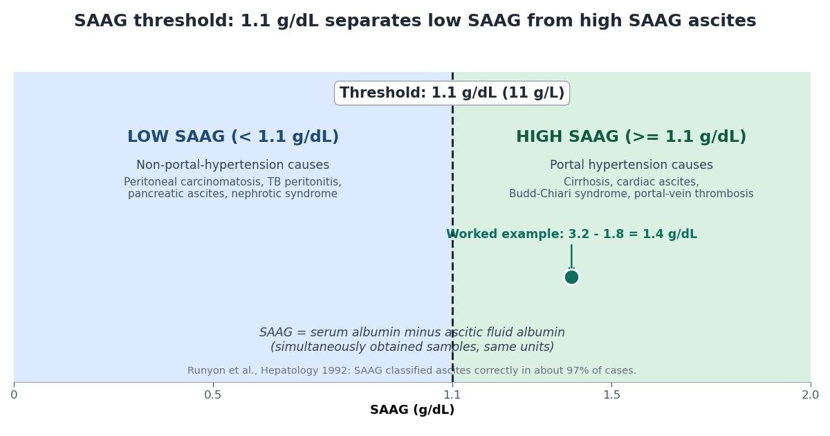
The gradient was introduced by Runyon and colleagues in a 1992 paper published in Hepatology, titled "The serum-ascites albumin gradient is superior to the exudate-transudate concept in the differential diagnosis of ascites." The study compared the new gradient against the older protein-based transudate/exudate classification and found that SAAG correctly classified the cause of ascites in about 97% of cases, substantially outperforming the older method. That result is the basis for the threshold the calculator above uses: a SAAG of 1.1 g/dL (11 g/L) or higher indicates a portal hypertension cause, and a SAAG below 1.1 g/dL indicates a non-portal-hypertension cause.
The 1.1 g/dL threshold
The entire clinical value of the SAAG is carried by a single number: 1.1 g/dL, equivalent to 11 g/L. This is a strict boundary. A SAAG of exactly 1.1 g/dL counts as a high SAAG and points to portal hypertension; a SAAG of 1.09 g/dL counts as a low SAAG and points away from it. The calculator applies this boundary exactly as published.
High SAAG (1.1 g/dL or higher): portal hypertension. A high gradient means the oncotic pressure of the blood is substantially higher than that of the ascitic fluid, which happens when hydrostatic pressure in the portal circulation forces fluid out of the hepatic sinusoids faster than albumin can follow. The classic causes are cirrhosis, which is by far the most common cause of high-SAAG ascites in most practice settings, cardiac ascites from right-sided heart failure, Budd-Chiari syndrome (obstruction of the hepatic venous outflow), and portal-vein thrombosis. All four share one mechanism: increased portal venous pressure.
Low SAAG (below 1.1 g/dL): non-portal-hypertension. A low gradient means the albumin concentrations in blood and fluid are relatively close, which happens when the ascites is produced by processes other than raised portal pressure. The causes Runyon's framework lists include peritoneal carcinomatosis (malignant ascites, where tumor implants increase peritoneal permeability and leak protein-rich fluid), tuberculous peritonitis (an inflammatory exudate), pancreatic ascites (from a pancreatic fistula or duct disruption, producing enzyme- and protein-rich fluid), and nephrotic syndrome (where massive urinary protein loss lowers serum albumin so severely that the gradient narrows). These are fundamentally different diseases from the portal hypertension group, and they require fundamentally different treatment.
| SAAG result | Interpretation | Typical causes |
|---|---|---|
| 1.1 g/dL or higher (11 g/L or higher) | High SAAG: portal hypertension cause | Cirrhosis, cardiac ascites, Budd-Chiari syndrome, portal-vein thrombosis |
| Below 1.1 g/dL (below 11 g/L) | Low SAAG: non-portal-hypertension cause | Peritoneal carcinomatosis, tuberculous peritonitis, pancreatic ascites, nephrotic syndrome |
How portal hypertension creates the gradient
The gradient is not a statistical trick; it has a direct physiological basis in the Starling forces that govern fluid movement across capillary walls. Fluid moves out of vessels when hydrostatic pressure pushes it and when the oncotic pressure of the surrounding fluid fails to hold it back, and it is reabsorbed when oncotic pressure dominates. In a healthy liver, the sinusoids are highly permeable, so plasma and hepatic lymph are nearly identical in protein content, and the albumin concentrations in blood and in any peritoneal fluid stay close together, keeping the gradient small.
Portal hypertension changes this balance. Raised pressure in the portal venous system increases hydrostatic pressure in the hepatic sinusoids, forcing large volumes of fluid out into the space of Disse and thence into the peritoneal cavity. This fluid is relatively protein-poor, because the high flow rate and the pressure gradient carry water and small solutes out faster than albumin can follow. At the same time, the diseased liver often synthesizes less albumin, so serum albumin falls. The result is a widening gap: serum albumin stays comparatively higher while ascitic fluid albumin falls, producing a large gradient. That is why a SAAG of 1.1 g/dL or more reflects portal hypertension so reliably, and why the gradient correlates with the portal pressure itself.
In the non-portal-hypertension causes, this mechanism is absent. In peritoneal carcinomatosis and tuberculous peritonitis, the fluid is produced by inflamed or tumor-infiltrated peritoneum that leaks protein freely, so ascitic fluid albumin stays high and the gradient stays narrow. In nephrotic syndrome, serum albumin is so low from urinary losses that there is little oncotic difference to maintain, again narrowing the gradient. In pancreatic ascites, the fluid is rich in pancreatic enzymes and protein from a ductal leak. Each of these keeps the two albumin values close together, producing a low SAAG.
Why the transudate/exudate concept was abandoned
Before 1992, ascites was classified by the total protein content of the ascitic fluid. Fluid with protein below 2.5 g/dL was called a transudate and attributed to cirrhosis; fluid with protein above 2.5 g/dL was called an exudate and attributed to malignancy or infection. This seemed biologically sensible, since transudates were thought to be low-protein filtrates and exudates protein-rich inflammatory fluids. The problem was that real patients did not respect the scheme.
The scheme's fatal flaw was diuretic therapy. Diuretics, the mainstay of treatment for cirrhotic ascites, concentrate the remaining ascitic fluid as water is removed, raising its protein content. A patient with straightforward cirrhosis on diuretics could therefore present with ascitic fluid protein above 2.5 g/dL, a pseudo-exudate that the old system misclassified as malignant or infectious. Conversely, some patients with peritoneal carcinomatosis presented with protein below the cutoff. The misclassification rate was substantial enough that the system actively misled treatment.
Runyon's insight was that the albumin gradient is largely immune to this problem. Diuretics raise both serum and ascitic fluid albumin concentrations in parallel, so the difference between them, the gradient, stays roughly constant even as the absolute values move. By comparing the fluid to the patient's own serum at the same moment, the gradient automatically corrects for the concentrating effect of treatment. The 1992 study showed that SAAG classified ascites correctly in about 97% of cases, compared with the substantially lower accuracy of the protein-based concept, and the transudate/exudate terminology for ascites was abandoned in favor of the gradient. Modern guidelines, including the 2021 AASLD update, use SAAG rather than total protein for the initial classification of ascites.
Getting the samples right
The SAAG is only as good as its two measurements, and three practical rules protect the calculation. First, the samples must be simultaneous. The serum sample and the ascitic fluid sample should be drawn at the same encounter, typically the blood drawn when the diagnostic paracentesis is performed. Albumin levels are not static: diuretic doses change, albumin infusions are given, large-volume paracentesis removes protein, and the underlying disease evolves. A serum albumin from Monday and an ascitic fluid albumin from Thursday describe two different physiological states, and their difference is not a SAAG. The 1.1 g/dL threshold and the 97% accuracy figure were validated on simultaneously drawn samples, so non-simultaneous sampling is the single most common way to get a wrong answer from a right formula.
Second, the units must match. Laboratories report albumin in either g/dL or g/L, and the two differ by a factor of ten: 1.1 g/dL equals 11 g/L. Mixing units, subtracting a g/L ascitic value from a g/dL serum value, produces a meaningless number. The calculator above applies one unit to both fields and converts between them on the standard factor of ten, but when reading laboratory reports manually, always confirm that both values use the same unit before subtracting.
Third, use albumin, not total protein. The gradient requires the albumin concentration specifically, measured in both serum and ascitic fluid. Total protein includes globulins and other proteins whose distribution does not track the oncotic gradient in the same way. Substituting total protein for albumin is a common error, particularly in settings still accustomed to the old transudate/exudate system, and it invalidates the interpretation.
A final practical point: the ascitic fluid albumin should not exceed the serum albumin in a valid measurement. Albumin in the peritoneal fluid comes from the plasma, so a fluid value higher than the serum value indicates a laboratory or sampling error, not a physiological state. The calculator flags this combination as an error rather than computing a negative gradient.
A worked example
Consider a patient whose laboratory reports a serum albumin of 3.2 g/dL and an ascitic fluid albumin of 1.8 g/dL, drawn at the same time. The SAAG is:
SAAG = 3.2 - 1.8 = 1.4 g/dL
Since 1.4 g/dL is at or above the 1.1 g/dL threshold, this is a high SAAG, pointing to a portal hypertension cause of the ascites. In most settings the leading candidate is cirrhosis, but cardiac ascites, Budd-Chiari syndrome, and portal-vein thrombosis all produce high SAAGs, and the gradient cannot distinguish among them. Further workup, typically including the ascitic fluid total protein, cell counts, and imaging, is needed to identify the specific disease.
In g/L units, the same patient would show a serum albumin of 32 g/L and an ascitic fluid albumin of 18 g/L. The SAAG is 32 - 18 = 14 g/L, which is at or above the 11 g/L threshold, the same conclusion. The calculator performs this unit conversion automatically; the interpretation is identical in either unit system.
Now consider the opposite case: serum albumin 2.8 g/dL and ascitic fluid albumin 2.2 g/dL. The SAAG is 2.8 - 2.2 = 0.6 g/dL, which is below 1.1 g/dL. This is a low SAAG, pointing away from portal hypertension and toward causes such as peritoneal carcinomatosis, tuberculous peritonitis, pancreatic ascites, or nephrotic syndrome. The workup now turns toward cytology, cultures, amylase, and renal function rather than portal pressure.
What SAAG cannot do
The SAAG is a classifier, not a diagnosis. A high SAAG tells the clinician that portal hypertension is driving the ascites, but it does not say whether the cause is cirrhosis, heart failure, Budd-Chiari syndrome, or portal-vein thrombosis. Distinguishing among these requires the rest of the ascitic fluid analysis and clinical evaluation. One useful refinement within the high SAAG group is the ascitic fluid total protein: a high SAAG with a total protein of 2.5 g/dL or higher suggests cardiac ascites rather than cirrhosis, because the failing heart produces a protein-rich transudate. The protein value that the old system misused as a classifier still has a role, but as a second step within the high SAAG category, not as the first branch of the workup.
A low SAAG is similarly only the start of the investigation. It rules out portal hypertension as the driver, but peritoneal carcinomatosis, tuberculous peritonitis, pancreatic ascites, and nephrotic syndrome are very different diseases, and each needs its own confirmation: cytology for malignancy, mycobacterial studies for tuberculosis, amylase for pancreatic leaks, and urinary protein quantification for nephrotic syndrome. The gradient narrows the field; it does not finish the job.
It is also worth stating plainly that the SAAG says nothing about the complications of ascites. It does not detect spontaneous bacterial peritonitis, which requires the ascitic fluid neutrophil count; it does not assess renal function or the risk of hepatorenal syndrome; and it does not measure the severity of the underlying liver disease, which is the domain of scores such as Child-Pugh and MELD. The gradient answers one question, the mechanism behind the fluid, and answers it well. Everything else requires the full clinical and laboratory picture.
Limitations
The SAAG has the limitations of the study it came from and of any single laboratory calculation. Runyon's 1992 study was a single-center study, and while its 97% accuracy has held up in subsequent practice and guideline endorsement, no test classifies every patient correctly. Mixed causes of ascites exist: a patient can have cirrhosis and peritoneal carcinomatosis simultaneously, and in such cases the gradient reflects the dominant physiology and may mislead if taken alone.
Treatment can also complicate interpretation. Although the gradient is more resistant to diuretic effects than the old protein classification, very large volume paracentesis with albumin replacement, intravenous albumin infusions, and severe intercurrent illness can all shift the two albumin values. The gradient is most reliable in the initial diagnostic workup of untreated or minimally treated ascites, which is exactly the setting guidelines recommend it for.
Finally, the threshold is a published convention, not a law of nature. A SAAG of 1.1 g/dL sits exactly on the boundary and is classified as high, but a patient with a gradient of 1.1 has less separation from the low group than a patient with a gradient of 2.0. Borderline results deserve the same caution as any borderline laboratory value: repeat the sampling if there is doubt, confirm the units, and interpret the number alongside the clinical picture rather than as an isolated verdict.
Key takeaways
- The serum-ascites albumin gradient is the serum albumin concentration minus the ascitic fluid albumin concentration, measured on simultaneously obtained samples in the same units.
- SAAG is calculated with one subtraction: serum albumin minus ascitic fluid albumin.
- A SAAG of 1.1 g/dL (11 g/L) or higher indicates that the ascites is caused by portal hypertension, with about 97% accuracy for distinguishing portal-hypertensive from non-portal-hypertensive ascites in the original Runyon study.
- A SAAG below 1.1 g/dL (11 g/L) indicates that the ascites is not caused by portal hypertension.
Frequently asked questions
What is the serum-ascites albumin gradient (SAAG)?
The serum-ascites albumin gradient is the serum albumin concentration minus the ascitic fluid albumin concentration, measured on simultaneously obtained samples in the same units. It reflects the oncotic pressure difference between blood and ascitic fluid and is used to classify ascites as caused by portal hypertension or by non-portal-hypertension disease. It replaced the older transudate/exudate classification based on ascitic fluid total protein.
How is SAAG calculated?
SAAG is calculated with one subtraction: serum albumin minus ascitic fluid albumin. Both samples must be drawn at the same time and reported in the same units, either g/dL or g/L. For example, a serum albumin of 3.2 g/dL and an ascitic fluid albumin of 1.8 g/dL gives a SAAG of 1.4 g/dL.
What does a SAAG of 1.1 g/dL or higher mean?
A SAAG of 1.1 g/dL (11 g/L) or higher indicates that the ascites is caused by portal hypertension, with about 97% accuracy for distinguishing portal-hypertensive from non-portal-hypertensive ascites in the original Runyon study. Portal-hypertension causes include cirrhosis, cardiac ascites, Budd-Chiari syndrome, and portal-vein thrombosis. The gradient itself does not identify which of these diseases is responsible.
What does a low SAAG (below 1.1 g/dL) mean?
A SAAG below 1.1 g/dL (11 g/L) indicates that the ascites is not caused by portal hypertension. Common causes of a low SAAG include peritoneal carcinomatosis (malignant ascites), tuberculous peritonitis, pancreatic ascites, and nephrotic syndrome. These conditions produce ascites through inflammation, infection, or low plasma oncotic pressure rather than raised portal pressure.
Why must the serum and ascitic fluid samples be drawn at the same time?
Albumin concentrations change with treatment and time. Diuretics, albumin infusions, paracentesis, and the natural course of the illness all move serum and ascitic fluid albumin levels, and a gradient computed from samples drawn on different days mixes two different physiological states. Simultaneous sampling ensures the gradient reflects a single moment of the patient’s physiology, which is what the 1.1 g/dL threshold and its 97% accuracy were validated against.
Why did SAAG replace the transudate/exudate classification of ascites?
The older system classified ascites as transudate or exudate based on ascitic fluid total protein, with 2.5 g/dL as the dividing line. It failed because diuretic therapy raises ascitic fluid protein and can convert a cirrhotic transudate into a pseudo-exudate, while malignant ascites can present with low protein. Runyon and colleagues showed in 1992 that the serum-ascites albumin gradient classified ascites correctly in about 97% of cases, far better than the protein-based concept, and major guidelines now recommend SAAG for the initial workup of ascites.
Medical disclaimer
This calculator is an educational tool that computes the serum-ascites albumin gradient exactly as published in Runyon et al., Hepatology 1992. It is not medical advice and is not a diagnosis. The SAAG classifies the mechanism behind ascites; it does not identify the specific disease, and it never replaces clinical judgment or a full diagnostic workup. All laboratory values should be obtained and interpreted by qualified clinicians, and new-onset ascites always requires prompt professional medical evaluation.
Sources
- Runyon BA, Montano AA, Akriviadis EA, Antillon MR, Irving MA, McHutchison JG. The serum-ascites albumin gradient is superior to the exudate-transudate concept in the differential diagnosis of ascites. Hepatology. 1992;15(6):982-996. doi:10.1002/hep.1840150603
- Biggins SW, Angeli P, Garcia-Tsao G, Gines P, Ling SC, Nadim MK, Wong F, Kim WR. Diagnosis, evaluation, and management of ascites, spontaneous bacterial peritonitis and hepatorenal syndrome: 2021 update by the American Association for the Study of Liver Diseases. Hepatology. 2021;74(2):e101-e131.