Light's Criteria Calculator: Exudate vs Transudate Pleural Effusion
In short: Apply Light's criteria to pleural fluid results: enter pleural and serum protein, pleural and serum LDH, and your lab's LDH upper limit to classify an effusion as exudate or transudate, with all three thresholds explained. Use the calculator above, then read the guide below to interpret your result and its limitations.
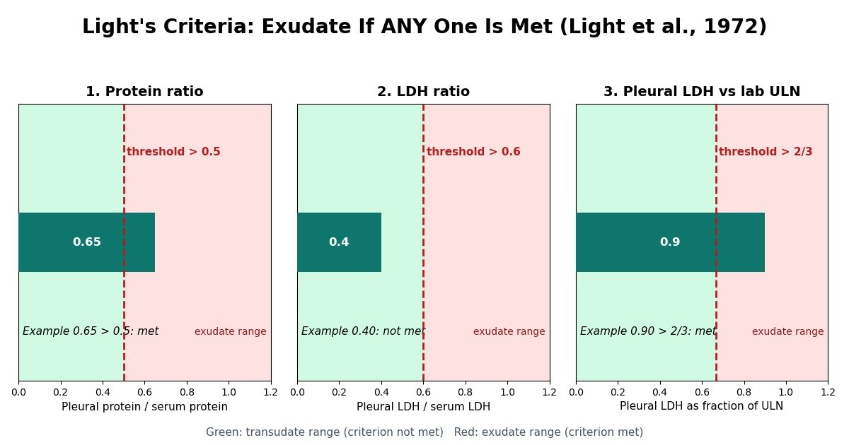
Light's criteria classify a pleural effusion as an exudate if ANY one of three tests is met: pleural fluid protein divided by serum protein above 0.5, pleural fluid LDH divided by serum LDH above 0.6, or pleural fluid LDH above two-thirds of the laboratory's upper limit of normal for serum LDH. Meet none of them and the effusion is a transudate. First described by Light and colleagues in 1972, the criteria remain the standard first step in pleural fluid analysis because they miss almost no exudates. Enter the five values below to apply all three checks.
Apply Light's criteria
All five values are required. Enter both protein values in the same unit, and all three LDH values in the same unit.
How to read your result
| Criterion | What the calculator checks | Threshold (strictly greater than) | If met |
|---|---|---|---|
| Protein ratio | Pleural fluid protein divided by serum protein | > 0.5 | Points toward exudate |
| LDH ratio | Pleural fluid LDH divided by serum LDH | > 0.6 | Points toward exudate |
| Pleural fluid LDH | Pleural fluid LDH against the lab's upper limit of normal | > 2/3 of the ULN | Points toward exudate |
The three checks use a parallel "or" rule: any single criterion met means the effusion is classified as an exudate, and this was the explicit design goal of the original 1972 paper, to maximise the identification of exudates. A transudate is defined by exclusion: all three criteria must fail for the effusion to be called a transudate. Note the thresholds are strictly "greater than". A protein ratio of exactly 0.5, an LDH ratio of exactly 0.6, or a pleural LDH exactly equal to two-thirds of the ULN does not meet the criterion; only values above the cutoffs count. The calculator applies this strict rule exactly, so borderline values on the wrong side of a threshold are reported as not met.
The three criteria, explained
1. Pleural fluid protein to serum protein ratio above 0.5
Protein is the oldest and most intuitive of the three markers. A healthy pleura is a tight barrier: protein leaks out of capillaries only slowly, so normal pleural fluid is low in protein relative to blood. When inflammation or malignancy injures the pleural membranes or blocks lymphatic drainage, protein pours into the pleural space and the fluid-to-serum protein ratio rises. A ratio above 0.5 is the single most used exudate flag, and in one later study effusions meeting both this criterion and the LDH threshold were considered conclusively exudative. Because this is a ratio, the absolute unit cancels out: g/dL and g/L give exactly the same answer as long as both values use the same unit.
2. Pleural fluid LDH to serum LDH ratio above 0.6
Lactate dehydrogenase is an intracellular enzyme released whenever cells break down. An inflamed or infected pleura, a growing tumour, or dying lung tissue spills LDH into the pleural space, so the pleural-to-serum LDH ratio reflects local tissue injury rather than barrier leakiness. The ratio above 0.6 catches exudates whose protein has not yet risen much, for example early parapneumonic effusions. Like the protein ratio, it is unitless: both values must simply be in the same unit.
3. Pleural fluid LDH above two-thirds of the serum LDH upper limit
The third criterion handles an edge case the ratios can miss: an exudate in a patient whose serum LDH is itself very high, which would depress the LDH ratio even though the pleural fluid is highly inflammatory. Comparing the absolute pleural LDH against the laboratory's reference range avoids that trap. The cutoff is two-thirds of the upper limit of normal for serum LDH, and it is the one criterion that depends on your laboratory's assay: different LDH assays produce different reference ranges, and platform differences have been shown to shift effusion classification, so you should always use the ULN printed on your own lab's report.
Why the exudate-transudate split matters
When fluid accumulates in the pleural space, the first clinical question is not "what is the exact disease" but "which family of diseases is this". A transudate forms when an imbalance of hydrostatic and oncotic pressures pushes fluid across an intact pleural membrane: the membrane is healthy and the problem is systemic, so the answer lies outside the chest. The classic transudative causes are heart failure, liver cirrhosis, and nephrotic syndrome, and treatment is aimed at the underlying condition, for example diuresis for heart failure or albumin management in cirrhosis. An exudate forms when the pleura itself is diseased: inflammation increases capillary leak, or lymphatic drainage is blocked. Here the answer is in the chest, and the workup shifts to the pleura itself, with Gram stain and cultures, cytology for malignancy, adenosine deaminase testing for tuberculosis, and pH and glucose measurement in suspected parapneumonic effusions.
This single split therefore halves the differential and decides the next step. More than sixty causes of pleural effusion are described, but four account for about three-quarters of cases: heart failure, cancer, pneumonia, and tuberculosis. The first of those four typically produces transudates, while the other three typically produce exudates, so getting the classification right steers the patient toward the correct three-quarters-or-one-quarter of the possibilities. Getting it wrong has a real cost: a transudate misclassified as an exudate can send a heart-failure patient for unnecessary pleural biopsy, while an exudate misclassified as a transudate can delay the diagnosis of malignancy or infection.
How accurate are Light's criteria?
The original description by Light, MacGregor, Luchsinger, and Ball in 1972 reported 99% sensitivity and 98% specificity for identifying exudates. Later studies have confirmed the first number but not the second: the ability of the criteria to rule out exudates has held up in multiple series and in a well-conducted meta-analysis, with sensitivity consistently near 100%, but real-world specificity is closer to 70%. In practice, 15% to 30% of true transudates are misclassified as exudates. The trade-off is deliberate: the criteria were designed with a parallel "or" rule specifically to maximise the identification of exudates, because missing an exudate (and therefore missing tuberculosis, malignancy, or empyema) was considered the more dangerous error.
Where do the false positives come from? The leading cause is diuretic therapy. As diuretics remove water from the body, protein and LDH in the pleural fluid become concentrated even though the membrane itself is unchanged, and a transudate that previously sat comfortably below the cutoffs creeps above them. This is why heart failure and cirrhosis, the two transudative conditions most often treated with long-term diuretics, dominate the misclassified group: in a European Respiratory Society study of 361 effusions, 35.4% of heart-failure-related and 33.9% of hepatic-hydrothorax-related effusions were misclassified as exudates by standard Light's criteria. More than half of misclassified transudates meet only one of the three criteria, with values sitting near the established threshold, which is a useful red flag: a single barely-positive criterion in a diuretic-treated patient deserves scepticism.
One further point in the criteria's favour: despite decades of proposed alternatives, Light's criteria remain the standard initial test, and they outperform clinical judgment alone for the transudate-exudate separation. A meta-analysis confirmed the cutoffs for the LDH and protein ratios, though it suggested that a pleural LDH above 45% of the upper limit of normal might be an even better single threshold than two-thirds.
When the result disagrees with the clinical picture
The criteria are a screening step, not a verdict. When Light's criteria label an effusion an exudate but everything about the patient says transudate (known heart failure, bilateral effusions, an elderly patient on diuretics), guidelines recommend rescue tests rather than an automatic pleural workup. The most studied is the serum-minus-pleural-fluid albumin gradient: a gradient above 1.2 g/dL indicates a transudate regardless of what Light's criteria say. Roth and colleagues introduced this cutoff in 1990, and in the European Respiratory Society study it correctly reclassified 70% of heart-failure false exudates and 56% of hepatic-hydrothorax false exudates, making it the most suitable parameter for cardiac false exudates.
A second gradient uses total protein instead of albumin: a serum-minus-pleural-fluid protein gradient above 3.1 g/dL indicates a transudate, and this was Light's own recommendation for sorting out discordant cases. In the same European study it reclassified 62% of heart-failure false exudates. For hepatic hydrothorax specifically, a pleural-fluid-to-serum albumin ratio below 0.6 correctly reclassified 56% of false exudates, slightly outperforming the albumin gradient in that subgroup. Other proposed refinements include pleural fluid NT-proBNP above 1500 pg/mL, which points strongly to a cardiac effusion, and pleural fluid cholesterol above 45 mg/dL as a single-test alternative with exudate accuracy comparable to Light's criteria.
The practical message is simple: when the classification and the patient disagree, trust the clinical picture first and reach for a gradient or NT-proBNP before scheduling an invasive procedure. A patient over 75 with bilateral effusions, confirmed gradients, and a high NT-proBNP almost certainly has a cardiac transudate no matter which single Light criterion was barely crossed.
Units, lab variation, and common pitfalls
Three practical mistakes account for most wrong answers from the calculator. First, mixing units: both protein values must share one unit and all three LDH values must share one unit, because the criteria are ratios and comparisons, not absolute numbers. The built-in unit selector for protein exists precisely for this reason; pick whichever unit your report uses and enter both values in it. Second, the wrong ULN: the two-thirds criterion is only as good as the upper limit of normal you type in. LDH assays are not standardised across laboratories, and a 2017 study in the Journal of Clinical Pathology showed that analytical platform variability alone can change effusion classification, so always take the ULN from the same laboratory report as the pleural LDH value. Third, over-reading borderline results: because diuretics and near-threshold values are the main sources of error, a result that hangs on a single criterion crossed by a small margin should be treated as uncertain, confirmed with a gradient test, and discussed with the treating clinician rather than acted on as a hard verdict.
Two final cautions. Light's criteria require a contemporaneous serum sample: protein and LDH drawn days apart from the thoracentesis are not what the criteria were validated on. And a traumatic tap that mixes blood into the pleural sample raises both protein and LDH artificially; visibly bloody fluid should be interpreted with that in mind.
Clinical source: Light RW, MacGregor MI, Luchsinger PC, Ball WC Jr. Pleural effusions: the diagnostic separation of transudates and exudates. Annals of Internal Medicine. 1972;77(4):507-513. doi:10.7326/0003-4819-77-4-507. Accuracy and rescue-test figures: Agrawal V, Doelken P, Sahn SA. Pleural fluid analysis. CMAJ. 2018;190(10):E291-5; Badiei A et al. From light to sound: ultrasound outshines Light's criteria. Respirology. 2024; Roth BJ et al. The serum-effusion albumin gradient in the evaluation of pleural effusions. Chest. 1990; European Respiratory Society false-exudate study, 2020.
Key takeaways
- The split halves the differential diagnosis.
- Light's criteria were deliberately designed to catch exudates, and they do that extremely well: the original 1972 report found 99% sensitivity and 98% specificity, and later studies confirm sensitivity near 100%.
- Diuretics concentrate protein and LDH in the pleural fluid as water is removed, pushing a true transudate over Light's thresholds.
- Apply one of the rescue tests recommended for this exact situation.