What the oxygenation index measures
The oxygenation index (OI) is a bedside index of oxygenation failure that relates the severity of hypoxaemia to the intensity of mechanical ventilatory support required to maintain it. In plain terms, it answers a more honest question than blood gas numbers alone: not just how low the arterial oxygen is, but how hard the ventilator has to work to keep it there.
The index combines three variables. FiO2 is the fraction of inspired oxygen the ventilator delivers, a number between 0.21 (room air) and 1.0 (pure oxygen). Mean airway pressure (mPaw, in cmH2O) is the average pressure applied to the airways over the respiratory cycle and captures the pressure cost of the current ventilator strategy, including PEEP and the inspiratory phase. PaO2 (in mmHg) is the arterial oxygen tension measured on a blood gas. Multiplying FiO2 by mean airway pressure, then by 100 and dividing by PaO2, yields a single number that rises when oxygenation worsens or when more pressure is needed to sustain a given oxygen level.
This construction is the index's key strength. A patient can have an identical PaO2 on two different days, yet be far sicker on the second day if reaching that PaO2 now requires substantially higher pressures and oxygen. The oxygenation index captures that difference, which is why intensive care teams use it to grade severity, track response to therapy and make referral decisions.
The formula and a worked example
The formula is:
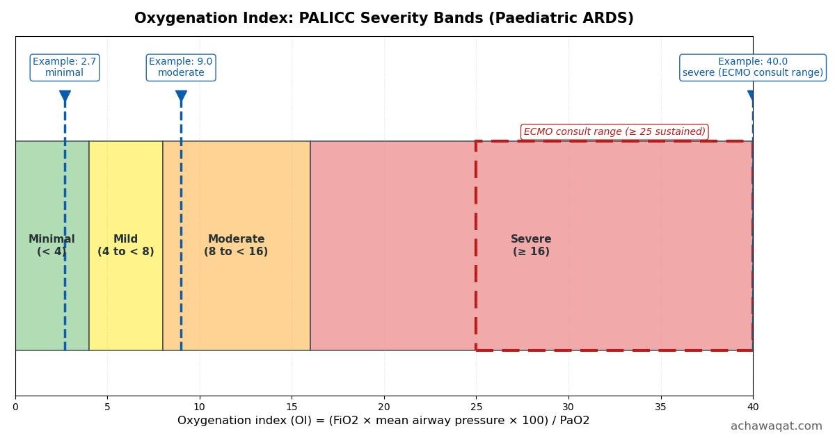
FiO2 must be entered as a fraction (for example 0.60 for 60% oxygen), mean airway pressure in cmH2O, and PaO2 in mmHg. Worked example: a ventilated child on FiO2 0.60, mean airway pressure 12 cmH2O, PaO2 80 mmHg gives OI = (0.60 × 12 × 100) / 80 = 720 / 80 = 9.0. Under the PALICC bands described below, an OI of 9.0 falls in the moderate range.
Because the formula divides by PaO2, the index is undefined when PaO2 is zero, which the calculator above treats as an input error rather than computing a value. Units must be consistent: entering mean airway pressure in the wrong unit, or PaO2 in kPa instead of mmHg, will silently produce a wrong answer, so confirm units before interpreting any result.
PALICC bands and the paediatric ARDS context
The Pediatric Acute Lung Injury Consensus Conference (PALICC) published its recommendations in 2015 (Khemani RG et al., Pediatr Crit Care Med 2015;16(5 Suppl 1):S23-40) and defined paediatric ARDS severity using the oxygenation index for invasively ventilated children. The PALICC cut-offs are: OI below 4, minimal or normal range; OI 4 to below 8, mild ARDS; OI 8 to below 16, moderate ARDS; and OI 16 or above, severe ARDS.
These bands matter because they standardise language. Saying a child has "severe paediatric ARDS by PALICC criteria" tells any clinician, in any centre, that the oxygenation index has reached 16 or more, which is far more precise than descriptive labels. The bands also underpin research: trials of ventilatory strategies and rescue therapies enrol patients by these thresholds, so a unit that documents OI in PALICC terms is speaking the same language as the evidence base.
For quick reference, the bands are:
| OI | PALICC severity |
|---|---|
| < 4 | Minimal or normal range |
| 4 to < 8 | Mild ARDS |
| 8 to < 16 | Moderate ARDS |
| ≥ 16 | Severe ARDS |
Note that the adult Berlin definition of ARDS grades severity with the P/F ratio rather than OI, so these bands apply to the paediatric context for which they were defined. In practice many clinicians also compute OI in adults, particularly when considering ECMO referral, but the formal PALICC bands belong to paediatric ARDS.
Oxygenation index versus the P/F ratio
The P/F ratio (PaO2 divided by FiO2) is the older and more widely quoted measure of oxygenation. Its limitation is structural: it ignores airway pressure entirely. Two patients can share a P/F ratio of 150 while one is on modest settings and the other requires a mean airway pressure above 20 cmH2O. The ratio treats them as equivalent; the oxygenation index does not, because mean airway pressure sits in its numerator.
This makes OI the more complete single number for a ventilated patient. A rising OI can signal deterioration even while the P/F ratio looks stable, because pressures may be climbing to hold the PaO2 steady. Conversely, a successful recruitment manoeuvre or a strategy change that maintains oxygenation at lower pressures will lower the OI, rewarding the gentler strategy. For trending a ventilated patient over days, OI is generally the more informative of the two.
The P/F ratio remains useful where ventilator data are unavailable or for quick comparisons, and the two indices usually move in opposite directions (OI rises as P/F falls). They are complementary descriptions of the same physiology, not competitors, but only OI accounts for the pressure cost.
The oxygen saturation index (OSI): a non-invasive alternative
The oxygen saturation index replaces the arterial blood gas with pulse oximetry:
PALICC also defined severity bands for OSI: below 5 indicates no or minimal ARDS, 5 to below 7.5 mild ARDS, 7.5 to below 12.3 moderate ARDS, and 12.3 or above severe ARDS. For example, FiO2 0.60, mean airway pressure 12 cmH2O and SpO2 92% give OSI = (0.60 × 12 × 100) / 92 = 7.8, which is moderate.
OSI's appeal is practical: it needs no arterial line and no blood gas, so it can be trended continuously at the bedside and used where arterial sampling is unavailable. Its weakness is the oximeter itself. Pulse oximetry is less reliable at very low saturations, saturations at or near 100% cannot discriminate further improvement (the flat top of the oxyhaemoglobin curve), and poor perfusion, motion or dyshaemoglobins can distort readings. Treat OSI as a useful screening and trending tool, and confirm important decisions with arterial blood gas based OI.
When clinicians use the oxygenation index
Three clinical uses dominate. The first is severity grading at diagnosis: once paediatric ARDS is recognised, the OI places it in the PALICC band, which sets expectations and guides the intensity of monitoring. The second is tracking response to therapy. An OI that falls after proning, a ventilator strategy change or a recruitment intervention suggests the intervention is working; an OI that climbs despite optimisation suggests the lung injury is progressing.
The third use is referral decision-making, particularly for extracorporeal membrane oxygenation (ECMO). Sustained OI values above 25 to 30 are commonly cited as a threshold for considering ECMO consultation, consistent with ELSO guideline practice. The word sustained matters: a single high reading during a transient event is not the same as persistent severe hypoxaemia on optimised ventilation. Referral also weighs the trajectory (improving or worsening), the reversibility of the underlying cause, contraindications, and the capability of the referring and receiving centres.
Because of these stakes, most units do not rely on a remembered number. Documenting the OI with its components (FiO2, mean airway pressure, PaO2, time) in the clinical record creates an auditable trail of severity over time, which is exactly what a referral centre will ask for.
Practical notes: steady state, suctioning and positioning
The oxygenation index is only as trustworthy as the moment it was measured. Several routine ICU events distort it transiently. Endotracheal suctioning typically drops saturations and PaO2 briefly, inflating the OI for minutes afterwards. Recent changes to ventilator settings, including PEEP adjustments and recruitment manoeuvres, mean the patient is not at a steady state and the index reflects a transition rather than the lung's true condition.
Position changes have similar effects: proning often improves oxygenation over hours, while the period immediately after turning can look worse. Agitation, ventilator asynchrony, circuit disconnections and changes in sedation all move the components independently of the underlying lung injury. The practical rule is simple: measure OI when the patient has been stable on unchanged settings for a reasonable period, note the time and context alongside the value, and trend it. A single value informs; a trend convinces.
Mean airway pressure itself deserves attention because it is a set and measured ventilator parameter rather than a physiological constant. Different ventilator modes report or achieve it differently, and switching modes can change mPaw without any change in the lung. When comparing OI values across days, confirm the ventilator mode and settings context as well as the number.
Limitations of the oxygenation index
OI is an index, not a diagnosis. It does not distinguish the cause of hypoxaemia: pneumonia, pulmonary oedema, atelectasis, pulmonary haemorrhage and intracardiac shunt can all raise it, and management differs completely between these. It is also influenced by factors outside the lung, including cardiac output, haemoglobin concentration and metabolic demand, which shift PaO2 and SpO2 without any change in lung injury.
The index assumes the inputs are accurate. A miscalibrated oxygen analyser, a damped arterial line, a venous sample mistaken for arterial blood, or a wrong unit all produce a confident-looking but wrong number. In paediatric practice the PALICC bands were derived for invasively ventilated children; applying them to non-invasive support or to adults is extrapolation, reasonable in experienced hands but not what the bands were validated for. Finally, OI says nothing about ventilator-induced lung injury risk beyond the pressure it includes: two identical OI values can carry different risks depending on tidal volumes and driving pressures.
A clinician tool, not a home triage test
The oxygenation index belongs at the bedside of a ventilated patient, interpreted by clinicians who can see the patient, the ventilator and the trend. It cannot be computed meaningfully at home: it requires a ventilator reporting mean airway pressure and an arterial blood gas or reliable oximetry, and its bands were defined for intensive care populations. If you are a parent or carer reading this page, the useful message is simpler: the care team tracks numbers like OI to grade how much support the lungs need, and questions about what the numbers mean for a specific patient belong in a conversation with that team, not with a calculator.
For clinicians, the index is one instrument among many. Use it to grade severity in PALICC terms, to trend response to therapy on comparable settings, and to document the case for escalation or referral. Pair it with the clinical examination, imaging, blood gases and the trajectory over hours, and it earns its place as one of the most practical numbers in ventilated paediatric care.