Modified Fisher Scale Calculator for Subarachnoid Haemorrhage
In short: Free modified Fisher scale calculator for subarachnoid haemorrhage: grade the admission CT by SAH thickness and intraventricular blood, with the approximate symptomatic vasospasm risk for each grade. Educational tool, not medical advice. Use the calculator above, then read the guide below to interpret your result and its limitations.
Grade the admission CT after subarachnoid haemorrhage with the modified Fisher scale. Two questions, SAH thickness and intraventricular blood, giving a grade from 0 to 4 with the approximate symptomatic vasospasm risk for each grade.
The calculator
Modified Fisher grade: Not yet calculated
CT findings: Select one option for each question
Approximate symptomatic vasospasm risk:
Please note: you selected intraventricular blood with no subarachnoid blood. The published scale defines grade 0 by the absence of SAH, and isolated IVH without any SAH falls outside what the scale was validated for, so this result needs a clinician's interpretation.
Please note: this is an educational aid. The risk figures are approximate cohort-level rates, not individual predictions. Subarachnoid haemorrhage is a medical emergency; any result here must be interpreted by the treating team.
What subarachnoid haemorrhage looks like on CT
Subarachnoid haemorrhage is bleeding into the space between the surface of the brain and the membranes (the meninges) that cover it. In most cases it follows the rupture of a small saccular aneurysm on one of the brain's arteries, although trauma and, less often, vascular malformations can cause it. The released blood spreads with the cerebrospinal fluid into the basal cisterns, the Sylvian fissures, the sulci over the brain's surface, and sometimes into the ventricles themselves.
A non-contrast CT scan of the head is the first test, because fresh blood appears bright (hyperdense) against the darker brain. The scan is most sensitive in the first day or two after the bleed; blood clears over the following days, so a CT done a week later can look deceptively clean. Radiologists describe where the blood sits and how much of it there is, and it was this pattern, visible on the admission scan, that Fisher and colleagues showed in 1980 could predict who would go on to develop vasospasm.
That link between the blood load and later vasospasm is the whole reason these scales exist. Vasospasm, the delayed narrowing of the brain's arteries, is one of the main causes of deterioration in the days after the initial bleed has been survived. A scale that grades the blood on the first CT gives the treating team an early, standardised sense of how closely to watch for it.
The original Fisher scale and its problem
Fisher, Kistler and Davis described the original scale in 1980. It had four grades: grade 1, no subarachnoid blood visible; grade 2, thin diffuse layers of blood under 1 mm; grade 3, localised clots or layers 1 mm or thicker; grade 4, intracerebral or intraventricular clot with diffuse or no subarachnoid blood.
The scale worked, but it had a structural weakness. Grade 4 was a mixed group: it lumped together patients whose main problem was blood inside the ventricles or the brain tissue, whatever the subarachnoid blood looked like. Intraventricular blood was treated as part of that leftover category rather than as a predictor in its own right, even though later evidence suggested it carried risk of its own.
In 2001, Claassen and colleagues showed that both cisternal blood and ventricular blood predicted delayed cerebral ischaemia independently of each other. That finding pointed to a cleaner design: instead of one axis of blood amount with a mixed fourth grade, use two axes, the thickness of the subarachnoid blood and the presence or absence of intraventricular blood, and let every combination have its own grade.
The modified Fisher scale: how to score it
The modified scale, published by Frontera and colleagues in Neurosurgery in 2006, does exactly that. Two findings from the admission CT decide the grade: how thick the subarachnoid blood is, and whether there is blood inside the ventricles. "Thin" means blood layers under 1 mm or focal deposits only. "Thick" means blood over 1 mm or filling the cisterns. Intraventricular haemorrhage means blood visible inside the ventricular system.
| Grade | Subarachnoid blood | Intraventricular blood | Description |
|---|---|---|---|
| 0 | None | Absent | No SAH, no IVH. |
| 1 | Thin or focal | Absent | Thin or focal SAH, no IVH. |
| 2 | Thin or focal | Present | Thin or focal SAH with IVH. |
| 3 | Thick or cisternal | Absent | Thick or cisternal SAH, no IVH. |
| 4 | Thick or cisternal | Present | Thick or cisternal SAH with IVH. |
The grade comes from the admission or early CT, because blood clears and a late scan underestimates the load. In practice, the radiologist's description is what you score from; the calculator above turns that description into the grade.
Why the modification matters
The modification is not cosmetic; it reflects how the risk actually behaves. Frontera's group studied 1,355 patients with subarachnoid haemorrhage, drawn from the placebo arms of four randomised trials, and 451 of them (33%) developed symptomatic vasospasm. After adjusting for other known predictors, each step up the modified scale raised the odds of vasospasm (adjusted odds ratio 1.28, 95% confidence interval 1.06 to 1.54). The original Fisher scale did not stay significant after the same adjustment.
The reason is the IVH axis. Ventricular blood predicts vasospasm independently of how much blood sits in the cisterns, so a thin bleed with IVH (grade 2) carries more risk than a thin bleed without it (grade 1), and a thick bleed with IVH (grade 4) carries more risk than a thick bleed without it (grade 3). The original scale could not show this, because its grade 4 mixed everything together. The modified scale's central insight is simple: two independent risks need two independent axes.
Understanding your result
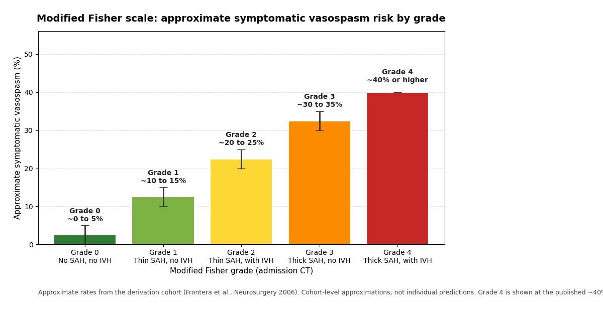
The approximate symptomatic vasospasm rates reported for each grade in the derivation cohort are shown in the table below. "Symptomatic vasospasm" means a new focal neurological deficit or a drop in consciousness that cannot be explained by something else, such as hydrocephalus, rebleeding, a seizure, or a metabolic disturbance. It is narrower than angiographic vasospasm (narrowing seen on imaging), which is more common and often causes no symptoms.
| Grade | Findings | Approximate symptomatic vasospasm risk |
|---|---|---|
| 0 | No SAH, no IVH | ~0 to 5% |
| 1 | Thin or focal SAH, no IVH | ~10 to 15% |
| 2 | Thin or focal SAH, with IVH | ~20 to 25% |
| 3 | Thick or cisternal SAH, no IVH | ~30 to 35% |
| 4 | Thick or cisternal SAH, with IVH | ~40% or higher |
These are approximations from one large cohort, and they describe groups, not individuals. A grade 4 result does not stamp a 40% personal probability on a patient; it means that among similar patients in the study, roughly four in ten developed symptomatic vasospasm. Other factors move individual risk up or down: older age, hypertension, a poor clinical grade at presentation, and early narrowing seen on angiography all add risk. Use the grade as a guide to vigilance, not as a fortune.
Why cisternal blood predicts vasospasm
The link is biological, not just statistical. Blood sitting in the subarachnoid space breaks down over days, releasing oxyhaemoglobin and other products that bathe the outside of the brain's arteries. These products disturb the vessel wall's normal balance: they promote constrictor signals such as endothelin, reduce the relaxing signal nitric oxide, and trigger inflammation around the vessel. The result is a prolonged spasm of the arterial muscle.
The timing follows the chemistry. Vasospasm typically develops between about day 4 and day 14 after the bleed, peaking around day 7, which matches when blood breakdown products are at their height. This is why the danger period comes days after the patient has survived the initial rupture, and why the grade on the admission CT is a forecast made in advance.
Narrowed arteries are not the whole story. Research has shown that delayed ischaemia can occur without demonstrable large-vessel narrowing, and that treatments which only reverse the narrowing have not always improved outcomes. Microcirculatory failure, spreading depolarisations, and early brain injury all contribute, which is why the field now often speaks of delayed cerebral ischaemia (DCI) rather than vasospasm alone. The modified Fisher scale predicts the clinical syndrome of symptomatic vasospasm as defined in 2006; it remains one of the best-validated early radiological predictors, but it is not a complete theory of DCI.
Monitoring for vasospasm and delayed cerebral ischaemia
Knowing the grade changes how closely the team watches, especially through the day 4 to 14 window. The cornerstone is the neurological examination: new weakness, a speech change, confusion, or a falling level of consciousness in that window is treated as possible vasospasm until another cause is found and excluded.
Transcranial Doppler ultrasound is the usual bedside monitor. Rising blood flow velocities in the brain's arteries, and the ratio of intracranial to extracranial velocities, suggest developing narrowing. CT perfusion or angiography can confirm it when the clinical picture is unclear.
Prevention and treatment are clinical decisions, but the standard measures are worth knowing. Nimodipine, a calcium-channel blocker, is given to reduce the risk of poor outcome related to vasospasm. Fluid balance is kept normal (euvolemic), because aggressive hypervolemia has not been shown to prevent ischaemia and carries its own risks. When delayed ischaemia is diagnosed, raising blood pressure can improve brain perfusion, and interventional options such as balloon angioplasty or vasodilator drugs delivered directly into the artery are available in specialist centres. None of this follows from the grade alone; the grade earns its keep by telling the team where on the risk spectrum this patient sits.
Worked examples
A CT shows a thin layer of blood in one Sylvian fissure and clear ventricles. Thin SAH with no IVH: grade 1, with an approximate symptomatic vasospasm risk of ~10 to 15%.
A CT shows thick blood filling the basal cisterns and blood layering in the ventricles. Thick SAH with IVH: grade 4, the highest grade, with an approximate risk of ~40% or higher.
A CT shows only a small focal deposit of blood but blood in the occipital horns of the ventricles. Thin SAH with IVH: grade 2, with an approximate risk of ~20 to 25%, higher than the grade 1 patient with the same thin bleed but clear ventricles. This is the IVH axis at work.
What the scale cannot do
The modified Fisher scale grades the scan, not the patient. Clinical severity is graded separately, with instruments such as the World Federation of Neurosurgical Societies (WFNS) grade or the Hunt and Hess grade; a patient can have a high Fisher grade and be clinically well, or a low grade and be deeply comatose. The two kinds of grading answer different questions.
It is a snapshot, not a forecast that updates itself. Blood clears, hydrocephalus can develop, and the patient's condition changes; the grade describes the admission CT and does not replace repeated assessment.
It does not diagnose the cause of the bleed, locate an aneurysm, or guide treatment by itself. A grade 4 does not mandate any particular intervention, and a grade 0 does not mean the patient is safe from every complication. Like every prediction tool, it informs clinical judgement; it never replaces it.
Key takeaways
- It measures the amount and distribution of blood on the admission CT scan after subarachnoid haemorrhage, as a predictor of symptomatic vasospasm.
- The original 1980 Fisher scale had four grades based on blood amount, with a mixed fourth grade for intraventricular or intracerebral clot.
- "Thin" means blood layers under 1 mm thick or focal deposits only.
- Studies found that ventricular blood predicts symptomatic vasospasm independently of how much blood sits in the cisterns, and that finding is the central insight of the modified scale.
Frequently asked questions
What does the modified Fisher scale measure?
It measures the amount and distribution of blood on the admission CT scan after subarachnoid haemorrhage, as a predictor of symptomatic vasospasm. Two findings decide the grade: how thick the subarachnoid blood is (none, thin or focal, or thick or cisternal) and whether there is blood inside the ventricles. The result is a grade from 0 to 4, with higher grades carrying a higher approximate risk of symptomatic vasospasm in the days after the bleed.
How is it different from the original Fisher scale?
The original 1980 Fisher scale had four grades based on blood amount, with a mixed fourth grade for intraventricular or intracerebral clot. It treated ventricular blood as part of that leftover category. The 2006 modified scale gives intraventricular blood its own axis: every SAH thickness is scored both with and without IVH, producing five grades. Because ventricular blood predicts vasospasm independently, grades 2 and 4 (with IVH) carry higher risk than grades 1 and 3, and the modified scale outperformed the original in a 1,355-patient study.
What counts as "thin" versus "thick" subarachnoid blood?
"Thin" means blood layers under 1 mm thick or focal deposits only. "Thick" means blood over 1 mm thick or filling the cisterns. The cutoff comes from the original Fisher work and was kept in the modified scale. In practice the radiologist's description of the admission CT is what you score from: a thin layer in the Sylvian fissure is thin, blood filling the basal cisterns is thick.
Why does blood inside the ventricles raise the vasospasm risk?
Studies found that ventricular blood predicts symptomatic vasospasm independently of how much blood sits in the cisterns, and that finding is the central insight of the modified scale. The likely reasons include a greater total blood load breaking down near the vessels and impaired cerebrospinal fluid circulation. Whatever the exact mechanism, the data are consistent: at any given SAH thickness, the grade with IVH carries a higher approximate risk than the grade without it.
What is the difference between symptomatic vasospasm and delayed cerebral ischaemia?
Symptomatic vasospasm, the endpoint the 2006 scale predicts, is a new focal neurological deficit or a drop in consciousness that cannot be explained by another cause such as hydrocephalus, rebleeding, a seizure or a metabolic disturbance. Delayed cerebral ischaemia is the broader modern concept: clinical deterioration or new infarction attributed to vasospasm after other causes are excluded. Some patients develop delayed ischaemia without demonstrable large-vessel narrowing, so vasospasm is one important mechanism of delayed cerebral ischaemia, not the whole story.
Can my Fisher grade be used to decide my treatment?
No. The grade is an educational and research instrument that guides how closely to watch for vasospasm; it does not prescribe any treatment. Management of subarachnoid haemorrhage weighs the grade alongside the clinical grade, the aneurysm treatment, other imaging, and the patient's whole condition. Only the treating team should make treatment decisions. This page does not diagnose subarachnoid haemorrhage and does not advise on treatment.
References and further reading
Medical disclaimer
This calculator is an educational aid only. It does not diagnose subarachnoid haemorrhage, does not replace clinical judgement, and does not advise on treatment. The modified Fisher scale is one instrument among many that clinicians use, and its result must be interpreted by a qualified clinician in the context of the full clinical picture, including the neurological examination and other imaging. Subarachnoid haemorrhage is a medical emergency: a sudden severe headache, collapse, or new neurological symptoms need emergency medical help immediately. Never make treatment decisions on the basis of a score alone.