Hunt-Hess Scale Calculator for Subarachnoid Haemorrhage
In short: Free Hunt-Hess scale calculator for subarachnoid haemorrhage: pick the clinical grade from 0 to 5, apply the systemic disease and vasospasm modifiers, and see the approximate surgical risk with honest framing. Educational tool, not medical advice. Use the calculator above, then read the guide below to interpret your result and its limitations.
Grade a subarachnoid haemorrhage on the Hunt-Hess scale: pick the clinical grade from 0 to 5 (including grade 1a), then apply the +1 modifier for serious systemic disease or severe vasospasm on angiography. The result shows the final grade with approximate historical surgical risk, framed honestly.
The calculator
Final Hunt-Hess grade: Not yet calculated
Select one clinical grade above
Please note: the +1 modifier convention applies to ruptured aneurysm grades 1 to 5. An unruptured aneurysm stays grade 0 regardless; serious systemic disease still raises operative risk for elective treatment, but it does not change the grade.
Please note: the surgical risk figure shown is an approximate historical figure from older surgical series. It is given to show the gradient across grades, not to predict any individual patient's outcome. Treatment has changed substantially since those series, and only the treating neurosurgical team can interpret a grade for a specific case.
Please note: this is an educational aid. It does not diagnose subarachnoid haemorrhage and does not guide treatment. Suspected subarachnoid haemorrhage is a medical emergency: seek immediate emergency care rather than using an online calculator.
What subarachnoid haemorrhage is
Subarachnoid haemorrhage is bleeding into the subarachnoid space, the fluid-filled gap between the arachnoid and pia mater membranes that surround the brain. Blood in this space irritates the meninges and raises intracranial pressure, and the sudden event can injure brain tissue directly. Most spontaneous cases come from a ruptured saccular (berry) aneurysm, a thin-walled outpouching of an artery, usually at a branch point in the circle of Willis or one of the major cerebral arteries. Other non-traumatic causes include arteriovenous malformations, and head injury is the most common cause of subarachnoid blood overall, though the Hunt-Hess scale was designed for aneurysmal bleeding.
The classic presentation is the thunderclap headache: a sudden, explosive headache that reaches maximum intensity within seconds to a minute, often described as the worst headache of the person's life. It is frequently accompanied by nausea and vomiting, photophobia, neck stiffness that develops over hours as the meninges become irritated, and sometimes a brief loss of consciousness at the moment of rupture. Focal neurological signs depend on where the aneurysm sits: a third cranial nerve palsy, with a dilated pupil and a drooping eyelid, classically points to a posterior communicating artery aneurysm pressing on the nerve. Seizures can occur, and bleeding into the eye (Terson syndrome) is a recognised sign of a severe bleed. In the worst cases the patient is comatose from the start because of acutely raised intracranial pressure or extensive brain injury.
Diagnosis starts with a non-contrast CT scan of the head, which is highly sensitive for subarachnoid blood in the first hours, though its sensitivity falls as the days pass and the blood clears. CT angiography then looks for the aneurysm itself. If the CT is negative but suspicion remains high, a lumbar puncture can detect blood breakdown products in the cerebrospinal fluid. Digital subtraction angiography remains the gold standard for detailed vascular anatomy and for planning treatment, whether that is surgical clipping of the aneurysm neck or endovascular coiling from inside the vessel. From the moment the diagnosis is made, the clinical team needs a quick, standardised way to describe how ill the patient is. That is what the Hunt-Hess scale provides.
Why clinicians grade subarachnoid haemorrhage
The question that led to the scale was a practical surgical one. In the 1960s, neurosurgeons knew that the risk of operating on a ruptured aneurysm depended heavily on the patient's condition at the time, and on whether the operation happened early or was delayed. William Hunt and Robert Hess, at Ohio State University, reviewed 275 consecutive cases of intracranial aneurysm treated over twelve years and asked whether the clinical picture at admission could predict surgical risk. Their answer, published in the Journal of Neurosurgery in 1968, was a five-grade classification: the worse the patient's neurological condition, the higher the surgical mortality and morbidity.
The scale was therefore born as a risk-stratification tool, not just a description. It let a surgeon estimate the danger of the operation from the bedside findings, and it shaped the debate about timing: operate early in good-grade patients before the aneurysm can rebleed, or wait in poor-grade patients until the brain recovers somewhat. Nearly sixty years later, that core insight still holds. Higher grades still mean worse outcomes, and the scale still serves as a common language: when a referral says "Hunt-Hess grade 3", every neurosurgeon knows roughly how ill that patient is. It also standardised how studies reported their patients, so that results from different centres and different eras could be compared.
The Hunt-Hess grades
The table below gives the grades as they are used today, with the wording kept close to the original descriptions.
| Grade | Clinical condition |
|---|---|
| 0 | Unruptured aneurysm. No subarachnoid haemorrhage has occurred. |
| 1 | Asymptomatic, or minimal headache and slight nuchal rigidity. |
| 1a | No acute meningeal or brain reaction, but a fixed neurological deficit is present. |
| 2 | Moderate to severe headache, nuchal rigidity, no neurological deficit other than cranial nerve palsy. |
| 3 | Drowsiness, confusion, or mild focal deficit. |
| 4 | Stupor, moderate to severe hemiparesis, possibly early decerebrate rigidity and vegetative disturbances. |
| 5 | Deep coma, decerebrate rigidity, moribund appearance. |
A note on provenance is important here, because an honest page should say what comes from 1968 and what came later. The original Hunt and Hess paper described grades 1 to 5. Grade 0, for an unruptured aneurysm, and grade 1a, for a patient with a fixed deficit but no acute meningeal reaction, were added in later use of the scale and are now standard in textbooks and teaching. They are included here because they are what clinicians actually use, but they were not part of the 1968 paper.
There is also the modifier rule, which the original paper stated plainly: for serious systemic disease, such as hypertension, diabetes, severe arteriosclerosis or chronic pulmonary disease, and for severe vasospasm seen on angiography, the patient is placed in the next less favourable grade. Each applies independently, so a patient with both is moved up two grades, with grade 5 as the ceiling. The calculator above applies this rule for you. One boundary needs stating: the modifier convention was written for ruptured aneurysm grades 1 to 5. An unruptured aneurysm stays grade 0 even when systemic disease is present, because there is no bleed to grade; the systemic disease still raises operative risk for elective treatment, which is a separate consideration.
Understanding your result: the mortality gradient
The figures below are the approximate surgical mortality and morbidity per grade commonly cited from the older surgical series. Read them as a gradient, not as predictions: what matters is how steeply risk rises from grade 1 to grade 5.
| Grade | Approximate historical surgical mortality/morbidity | What the gradient means |
|---|---|---|
| 1 | 0 to 5% | Low single digits in the historical series; the safest surgical group. |
| 2 | About 5 to 10% | Roughly one in ten to one in twenty; still a good surgical grade. |
| 3 | About 15 to 20% | Roughly one in five to one in seven; the middle ground where risk becomes substantial. |
| 4 | About 40% | Roughly two in five; poor-grade surgery carried very high risk. |
| 5 | 70% or more | Seven in ten or higher; surgery in this group was historically perilous. |
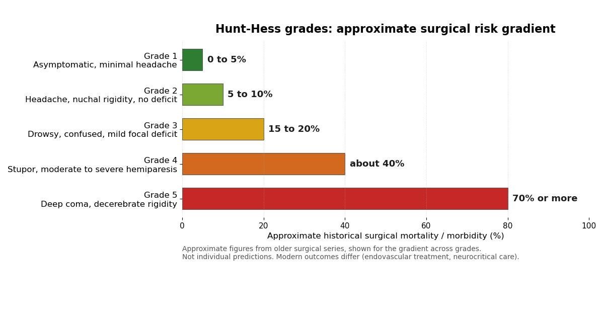
Honest framing matters more here than on most calculator pages, because these numbers look precise and they are not. They come from surgical series of the 1960s and 1970s, an era before endovascular coiling, before modern neurocritical care, before routine vasospasm prevention with nimodipine, and before the intensive monitoring that poor-grade patients receive today. Modern outcomes are better than these figures suggest, and they vary enormously between centres. The numbers also describe groups, not people: a grade 3 patient with a small anterior communicating artery aneurysm and no other problems is in a different position from a grade 3 patient who is elderly with severe heart disease. Use the table to understand why the grade matters, never to predict what will happen to an individual.
What the grade tends to mean for management
The grade shapes, but does not dictate, the plan. What follows is a general educational picture of how the grades are usually handled; real decisions are individualised by the neurosurgical team using the full clinical picture and imaging.
Grades 1 and 2 are good surgical grades. The usual priority is to secure the aneurysm early, by surgical clipping or endovascular coiling, because the greatest early danger is rebleeding, which carries a very high mortality. Early securing lets the team manage blood pressure and start vasospasm prevention without the aneurysm hanging over every decision. Grade 3 patients are often still treated early, but with a clear-eyed view of the higher risk; they need close neurological monitoring in a neurosurgical intensive care setting, because deterioration can be swift.
Grades 4 and 5 are poor grades, and here practice is more cautious. Operating on a deeply comatose, unstable brain adds a major insult to an already injured one, so the common approach is to stabilise first: manage intracranial pressure, support ventilation and circulation, prevent and treat vasospasm, and watch for hydrocephalus, which is common after subarachnoid haemorrhage and is treated with cerebrospinal fluid drainage. Intervention on the aneurysm itself may be deferred until the patient improves to a better grade, though emergency surgery is still done when there is a life-threatening mass lesion such as a large intracerebral haematoma that needs evacuating.
Across all grades, certain elements of care are constant. Nimodipine, a calcium channel blocker, is given to reduce the risk of delayed cerebral ischaemia from vasospasm. Blood pressure is controlled carefully before the aneurysm is secured, because hypertension promotes rebleeding, and managed differently afterwards. The days from roughly day 4 to day 14 after the bleed are the vasospasm window, when the team watches for new neurological deficits and uses transcranial Doppler and clinical examination to catch vasospasm early. Seizure prophylaxis is considered in selected patients, and hydrocephalus is watched for throughout. The grade is one input to all of this, alongside the CT findings, the aneurysm's size and location, the patient's age and other conditions, and often a WFNS grade as well.
Related scales: WFNS and Fisher
Hunt-Hess is not the only grading system, and modern practice usually uses more than one, because each scale answers a different question. The World Federation of Neurosurgical Societies (WFNS) scale was introduced in 1988 by Teasdale and colleagues precisely because Hunt-Hess relies on subjective descriptions. Terms like drowsiness, stupor and mild focal deficit mean slightly different things to different observers, and studies have shown meaningful interobserver variability, especially around the grade 2 to 3 boundary. The WFNS scale replaces the descriptions with the Glasgow Coma Scale plus the presence or absence of a major focal deficit (aphasia or hemiparesis): grade I is GCS 15 with no deficit, grade II is GCS 13 to 14 with no deficit, grade III is GCS 13 to 14 with a deficit, grade IV is GCS 7 to 12, and grade V is GCS 3 to 6. It is more reproducible, and many centres now report it as the primary clinical grade.
The Fisher scale answers a different question again: not how ill the patient looks, but how much blood is on the CT scan, because the amount and distribution of subarachnoid blood predicts vasospasm. Fisher, Kistler and Davis described it in 1980: grade 1 is no blood detected, grade 2 is diffuse thin layers under 1 mm, grade 3 is localised clot or layers over 1 mm, and grade 4 is intracerebral or intraventricular clot. The modified Fisher scale (Frontera and colleagues, 2006) refined it by giving intraventricular blood its own weight, since blood in the ventricles independently raises vasospasm risk. A patient can therefore have a low Hunt-Hess grade and a high Fisher grade, meaning they look well now but carry a high vasospasm risk, which is exactly why the scales are used together rather than as alternatives.
Limitations of the Hunt-Hess scale
An honest account of the scale includes its weaknesses. The main one is subjectivity: the boundary between drowsiness and stupor, or between a mild and moderate focal deficit, is a judgement call, and different clinicians can assign different grades to the same patient. It was also designed around surgical risk in an era when surgery was the only way to secure an aneurysm; endovascular coiling has changed the risk calculus for many aneurysms. The scale says nothing about vasospasm risk, which is why the Fisher scale exists, and nothing about the aneurysm's anatomy, which often drives the choice between clipping and coiling. It does not diagnose subarachnoid haemorrhage, and it was never meant to: it assumes the diagnosis is established. Finally, the historical outcome figures attached to it predate modern care and should not be quoted as current prognosis. None of this makes the scale useless; it makes it one instrument among several, to be used with its limits understood.
Worked examples
A few worked examples show how the grade and the modifiers fit together.
Example one. A 45-year-old arrives with a sudden severe headache and a stiff neck after a confirmed subarachnoid haemorrhage. She is alert, talking normally, with no weakness and no cranial nerve signs. That matches grade 2: moderate to severe headache, nuchal rigidity, no neurological deficit other than cranial nerve palsy. She has no serious systemic disease and the angiogram shows no severe vasospasm, so no modifiers apply. Final grade: 2, with an approximate historical surgical risk of about 5 to 10 percent.
Example two. A 62-year-old man is drowsy and confused with a mild weakness of one side: grade 3. He has long-standing diabetes and hypertension, which is serious systemic disease, and the angiogram shows severe vasospasm. Each adds one grade, so grade 3 moves up two to grade 5, the ceiling. Final grade: 5, with an approximate historical surgical risk of 70 percent or more. The modifiers have honestly captured how much worse his position is than the base grade 3 alone.
Example three. A 50-year-old woman has an aneurysm found incidentally on imaging done for another reason. It has never bled: grade 0. She has hypertension, which counts as serious systemic disease. The modifier rule applies to ruptured grades 1 to 5, so her grade stays 0, and the calculator says so plainly. Her surgical risk is the risk of elective aneurysm treatment, which varies by case and is not what the historical table describes.
Key takeaways
- The Hunt-Hess scale grades how ill a patient is after a subarachnoid haemorrhage from a ruptured brain aneurysm, from grade 1 (asymptomatic or minimal headache) to grade 5 (deep coma).
- The grade moves up one level if the patient has a serious systemic disease, such as hypertension, diabetes, severe arteriosclerosis or chronic pulmonary disease, or if there is severe vasospasm seen on angiography.
- Grade 0 means an unruptured aneurysm: no subarachnoid haemorrhage has occurred.
- The Hunt-Hess scale grades subarachnoid haemorrhage using clinical descriptions such as drowsiness, stupor and hemiparesis, which different observers can interpret differently.
Frequently asked questions
What is the Hunt-Hess scale used for?
The Hunt-Hess scale grades how ill a patient is after a subarachnoid haemorrhage from a ruptured brain aneurysm, from grade 1 (asymptomatic or minimal headache) to grade 5 (deep coma). Hunt and Hess introduced it in 1968 to estimate surgical risk: the higher the grade, the higher the surgical mortality and morbidity. It gives the whole team a common language for describing the patient's condition and helps guide decisions about the timing and risks of securing the aneurysm.
When does the Hunt-Hess grade go up by one?
The grade moves up one level if the patient has a serious systemic disease, such as hypertension, diabetes, severe arteriosclerosis or chronic pulmonary disease, or if there is severe vasospasm seen on angiography. Each applies independently, so both together add two, with the grade capped at 5. This calculator applies both modifiers and shows the final grade, or explains why the grade stays the same.
What are grade 0 and grade 1a?
Grade 0 means an unruptured aneurysm: no subarachnoid haemorrhage has occurred. Grade 1a means there is no acute meningeal or brain reaction, but the patient has a fixed neurological deficit. Both were added in later use of the scale; the original 1968 paper described only grades 1 to 5. The +1 modifier convention applies to the ruptured grades 1 to 5, so an unruptured aneurysm stays grade 0 regardless of other conditions.
What is the difference between the Hunt-Hess scale and the WFNS scale?
The Hunt-Hess scale grades subarachnoid haemorrhage using clinical descriptions such as drowsiness, stupor and hemiparesis, which different observers can interpret differently. The World Federation of Neurosurgical Societies (WFNS) scale, introduced in 1988, was designed to be more objective: it grades patients using the Glasgow Coma Scale together with the presence or absence of a major focal deficit. Many centres now use the WFNS scale alongside or instead of Hunt-Hess, and use the Fisher scale on the CT scan to estimate vasospasm risk.
Do the mortality figures on this page predict what will happen to an individual patient?
No. The figures shown are approximate surgical mortality and morbidity from historical surgical series, and they are presented so you can see the gradient across grades, not to predict any one person's outcome. Treatment has changed substantially since those series, with endovascular coiling, modern intensive care and vasospasm prevention, so current outcomes differ. An individual's prognosis depends on many factors beyond the grade, and only the treating neurosurgical team can interpret the result for a specific case.
Can this calculator be used to make treatment decisions?
No. This calculator is an educational aid for understanding the scale, not a decision tool. Subarachnoid haemorrhage is a medical emergency: anyone with a sudden severe headache or other suspicious symptoms needs immediate emergency assessment, not an online calculator. Decisions about securing an aneurysm, timing of surgery, and intensive care are made by a neurosurgical team using the full clinical picture, imaging and often several grading scales together.
References and further reading
Medical disclaimer
This calculator is an educational aid only. It does not diagnose subarachnoid haemorrhage, does not replace clinical judgement, and does not advise on treatment. Subarachnoid haemorrhage is a medical emergency: a sudden severe headache, collapse, or sudden neurological symptoms need immediate emergency care. The Hunt-Hess grade is one instrument among several that neurosurgical teams use, and its result must be interpreted by qualified clinicians in the context of the full clinical picture and imaging. The historical risk figures on this page describe older surgical series and must not be used to predict any individual's outcome. Never delay seeking emergency care because of anything on this page.