What the ICH Score is
Spontaneous intracerebral hemorrhage, bleeding directly into the brain tissue without trauma, accounts for roughly 10 to 15 percent of all strokes yet carries a far higher risk of death and disability than ischemic stroke or subarachnoid hemorrhage. In the cohort used to develop this scale, 45 percent of patients had died within 30 days. Because the early hours after a bleed demand rapid, high-stakes decisions, clinicians have long wanted a simple, standard way to describe how sick a patient with intracerebral hemorrhage is at presentation, similar to the Glasgow Coma Scale for head injury or the Hunt-Hess scale for aneurysmal subarachnoid hemorrhage.
The ICH Score, published by J. Claude Hemphill III and colleagues in Stroke in 2001, fills that gap. It combines five findings that are each independently associated with 30-day mortality into a single 0 to 6 score: the Glasgow Coma Scale on presentation, the hematoma volume on the initial CT, the presence of intraventricular hemorrhage, whether the bleed started below the tentorium, and whether the patient is 80 years or older. The score was designed so that any clinician, without special training in stroke neurology or statistics, can compute it at the bedside within minutes of seeing the first CT scan.
The developers built the scale from the records of 161 patients who presented with acute nontraumatic intracerebral hemorrhage to two University of California, San Francisco hospitals between January 1997 and December 1998; complete data were available for 152 of them. Logistic regression identified the five independent predictors of 30-day death, and each was weighted by the strength of its association with outcome. The Glasgow Coma Scale, the strongest predictor, received up to 2 points; each of the other four received 1 point.
The five components and their points
| Component | Finding | Points |
|---|---|---|
| Glasgow Coma Scale (on presentation or after resuscitation) | 13 to 15 | 0 |
| 5 to 12 | 1 | |
| 3 to 4 | 2 | |
| ICH volume on initial CT (ABC/2 method) | Less than 30 cm3 | 0 |
| 30 cm3 or more | 1 | |
| Intraventricular hemorrhage (any blood in the ventricles on initial CT) | Absent | 0 |
| Present | 1 | |
| Infratentorial origin (bleed starting in the cerebellum or pons) | No (supratentorial) | 0 |
| Yes | 1 | |
| Age | Younger than 80 years | 0 |
| 80 years or older | 1 | |
| Total ICH Score | 0 to 6 | |
Glasgow Coma Scale
The GCS sums eye opening (1 to 4), verbal response (1 to 5), and motor response (1 to 6) into a total from 3 to 15. It is the single strongest element of the ICH Score, which is why it alone can contribute 2 points. In the derivation cohort, only 1 of 35 patients presenting with a GCS of 3 or 4 survived to 30 days, while 29 of 57 patients with a GCS of 5 to 12 died, compared with 5 of 60 patients with a GCS of 13 to 15. Unusually among hemorrhage prediction models, the authors split the scale into three bands rather than two, because patients with scores of 3 or 4 did dramatically worse than those with higher scores regardless of other factors. Use the GCS recorded at the time of transfer from the emergency department, after resuscitation; a GCS depressed by sedation or intubation rather than the bleed itself will overstate the score.
ICH volume of 30 cm3 or more
Hematoma volume is one of the most robust predictors of outcome after intracerebral hemorrhage across many studies. The authors dichotomized it at 30 cm3 because that value marked the cut point for increased mortality in their cohort, is easy to remember, and matched volume thresholds used in earlier models. Adding extra points for very large hematomas, for example above 60 cm3, did not improve the accuracy of the scale, so the volume component stays binary. Note a quirk of the original data: no patient with an infratentorial bleed had a hematoma of 30 cm3 or more, which is one reason a score of 6 never occurred in the derivation cohort.
Intraventricular hemorrhage
Any blood visible in the cerebral ventricles on the initial CT earns 1 point. In the derivation cohort, 66 percent of patients with IVH died within 30 days versus 19 percent without it. IVH was the weakest of the five independent predictors and only borderline significant in the multivariable model, but it was retained because its association with outcome was consistent and it is simple to assess.
Infratentorial origin
Bleeds starting in the cerebellum or pons earn 1 point; supratentorial bleeds, including basal ganglia, thalamic, and lobar hemorrhages, earn 0. The posterior fossa leaves little room for an expanding hematoma, so even small bleeds there can compress the brainstem. Interestingly, volume was not an independent predictor of outcome within the infratentorial subgroup, probably because location dominates size when the bleed sits in the brainstem or cerebellum.
Age 80 years or older
Patients aged 80 or above earn 1 point. The authors dichotomized age at 80 because younger age bands showed no association with 30-day mortality in their data; only the 80-plus group stood apart, with 67 percent 30-day mortality versus 39 percent in younger patients. The single point reflects a modest but real independent contribution after accounting for the other four components.
How to estimate ICH volume with the ABC/2 method
Most clinicians do not have planimetric software at the bedside, so the ICH Score uses the ABC/2 ellipsoid approximation described by Kothari and colleagues in 1996. The method treats the hematoma as an ellipsoid and needs only three measurements from the non-contrast head CT:
- A: the greatest hemorrhage diameter, in centimeters, on the axial slice where the bleed appears largest.
- B: the diameter perpendicular to A, in centimeters, on that same slice.
- C: the approximate number of axial slices on which any hemorrhage is visible, multiplied by the slice thickness in centimeters. If the bleed appears on 6 slices cut at 5 mm thickness, C is 3.0 cm. A common refinement is to compare each slice with the largest one and count slices showing more than about 75 percent of that area as full slices, but the original method simply multiplies slice count by thickness.
The estimated volume is then (A x B x C) / 2 cubic centimeters. A worked example: A = 4.0 cm, B = 3.0 cm, and hemorrhage visible on 5 slices at 0.5 cm thickness, so C = 2.5 cm. The volume is (4.0 x 3.0 x 2.5) / 2 = 15 cm3, which is below the 30 cm3 threshold and earns 0 points. If instead the bleed measured 5.0 by 4.0 cm across 4.0 cm of slices, the volume would be (5.0 x 4.0 x 4.0) / 2 = 40 cm3, earning 1 point. The ABC/2 estimate correlates well with computer-measured volumes for roughly ellipsoid bleeds but becomes less accurate for very irregular or multilobed hematomas, a limitation worth remembering when the volume sits close to the 30 cm3 cut point.
What each score meant in the original cohort
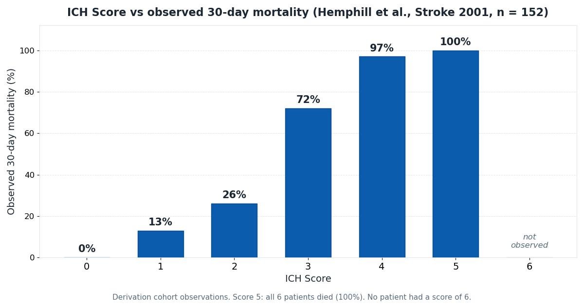
The table below shows the 30-day mortality actually observed for each ICH Score in the 152-patient derivation cohort. These are cohort observations, not individual predictions: they describe what happened to similar patients in one study, not what will happen to the patient in front of you.
| ICH Score | Observed 30-day mortality | What the original paper reported |
|---|---|---|
| 0 | 0% | All 26 patients survived |
| 1 | 13% | As reported in the paper |
| 2 | 26% | As reported in the paper |
| 3 | 72% | As reported in the paper |
| 4 | 97% | As reported in the paper |
| 5 | 100% | All 6 patients died |
| 6 | No observed data | No patient in the cohort had a score of 6 |
Mortality rose steadily with the score, a trend that was statistically significant both overall and within the supratentorial and infratentorial subgroups. That consistency across subgroups is what makes the ICH Score usable for all intracerebral hemorrhage patients rather than only a subset.
Validation in later cohorts
A score is only as useful as its performance outside the hospital where it was born. The most important validation came from Hemphill's own group: a prospective study of 243 patients admitted with acute intracerebral hemorrhage between June 2001 and May 2004 found that the ICH Score accurately stratified 12-month functional outcome measured with the modified Rankin Scale, at several cut points along that scale. This extended the score beyond its original 30-day mortality endpoint to long-term function, which matters more to patients and families. The study also documented something hopeful: many patients kept improving across the first year, with gains continuing even after six months, a reminder that early scores capture a moment, not a destiny.
Independent groups have tested the score as well. A large study of more than 1,200 patients with intracerebral hemorrhage, including patients on oral anticoagulants, confirmed that the original ICH Score stratified prognosis and that adding anticoagulant use did not improve its performance. Early independent testing, such as the report by Fernandes and colleagues in 2002, likewise supported the score's discriminative ability. No grading scale is perfect, but the ICH Score has held up across centers, countries, and decades, which is why it remains the most widely used bedside scale for this disease.
Using the score in practice
In the emergency department, the ICH Score earns its keep as a communication tool. Saying "ICH Score 4" conveys, in three syllables, a GCS in the low range or a large complicated bleed in an older patient, plus the knowledge that roughly 97 of 100 similar patients in the original cohort died within a month. That shared language helps emergency physicians, neurologists, neurosurgeons, intensivists, and families start from the same facts. Researchers use the score to stratify trial enrollment so that treatment arms contain comparable patients, one of the purposes the authors explicitly intended.
For families, the score can anchor an honest conversation, but it must be translated carefully. A useful framing is: "Among 100 patients who looked like this on arrival in the original study, about this many had died within a month." Pair the number with what the score cannot say: whether this particular patient will be among the survivors, what quality of life survivors had, and how modern care, which has changed since 1997 to 1998, might shift the odds. The 2022 American Heart Association and American Stroke Association guideline for spontaneous intracerebral hemorrhage emphasizes organized, aggressive early care, including blood pressure control and reversal of anticoagulation, and the broader stroke literature cautions that prognostic scores should supplement rather than replace clinical judgment.
Above all, the score must not become a shortcut to giving up. Because patients with devastating early scores sometimes survive with meaningful recovery, and because withdrawing care early guarantees the predicted death, guidelines and ethicists warn against basing early do-not-resuscitate orders or withdrawal decisions on a score alone. The original authors made the same point in their own way: they argued the scale should standardize assessment and communication, not dictate who gets treated.
Limitations
Every limitation below is a reason to treat the score as one input among many, not a verdict.
- Small, single-center, retrospective derivation. The score came from 152 patients at two hospitals in one city, treated in 1997 and 1998. Practice has changed since then, and small cohorts produce unstable estimates, especially at the extremes: the 100% mortality for score 5 rests on just 6 patients.
- No observed data for score 6. As discussed above, the highest score is an extrapolation, not a measurement.
- Withdrawal-of-care bias. Patients in whom life-sustaining treatment was withdrawn in the first days were included in the analysis. If high scores prompted earlier withdrawal, the observed mortality at high scores partly reflects decisions, not only biology, creating a self-fulfilling prophecy that later users of the score can perpetuate.
- Mortality is not function. The original endpoint was death at 30 days. Families usually care about what life looks like for survivors: independence, cognition, and disability. The 2009 prospective validation helps here by linking the score to 12-month functional outcome, but the famous 0-to-100 percentages describe death, not disability.
- Volume is an estimate. ABC/2 approximates irregular hematomas imperfectly, and a volume near the 30 cm3 cut point can flip the score by a point on measurement variation alone.
- GCS can mislead. Intubation, sedation, seizures, or intoxication can depress the GCS for reasons unrelated to the bleed, inflating the score. The scale asks for the post-resuscitation examination precisely to reduce this noise.
- It ignores much else. Hydrocephalus, hematoma expansion, blood pressure, glucose, anticoagulant use, and baseline frailty all influence outcome and none appear in the score. A low score does not guarantee safety, and a high score does not make treatment futile.