A-a Oxygen Gradient Calculator
In short: Free A-a oxygen gradient calculator: PAO2 from the alveolar gas equation, alveolar-arterial gradient, age-based expected gradient estimate, and what a widened or normal gradient means. Includes worked examples, physiology explainers, and FAQs. Use the calculator above, then read the guide below to interpret your result and its limitations.
Alveolar-arterial oxygen gradient from the alveolar gas equation. Enter the arterial blood gas values below; the calculator estimates alveolar oxygen (PAO2), computes the gradient (PAO2 minus PaO2), and, on room air, compares it against the age-based expected estimate. Physiology verified against StatPearls (NCBI Bookshelf) on 2026-10-05. Medically reviewed by Dr. Taimoor Asghar.
Calculator
0.21 for room air, 1.0 for 100% oxygen. Enter as a decimal, not a percentage.
Default 0.8 (steady-state average). Usually 0.7 to 1.0 depending on diet and metabolic state.
760 mmHg at sea level. Lower it at altitude.
What the A-a gradient measures
The alveolar-arterial (A-a) oxygen gradient measures how efficiently oxygen moves from the air sacs of the lungs into the arterial blood. It is the difference between the partial pressure of oxygen in the alveoli (PAO2), which we estimate with the alveolar gas equation, and the partial pressure of oxygen dissolved in arterial blood (PaO2), which we measure with an arterial blood gas (ABG): A-a gradient = PAO2 minus PaO2. In a perfect lung every molecule of oxygen that reached an alveolus would cross into the capillary beside it, and the gradient would be zero. Real lungs are not perfect. Even in healthy young adults, ventilation and blood flow are not matched identically across all lung regions: the bases receive relatively more blood flow than ventilation while the apices receive relatively more ventilation than blood flow. This physiologic ventilation-perfusion mismatch means a small gap between alveolar and arterial oxygen is normal.
The A-a gradient therefore answers a different question than the PaO2 alone. The PaO2 tells you that oxygen is low. The gradient tells you where to look for the reason: a widened gradient means the problem lies in gas exchange across the alveolar-capillary membrane, while a normal gradient points toward causes outside that membrane, such as breathing too little (hypoventilation) or breathing air with less oxygen in it. That single distinction is why the calculation appears in virtually every hypoxemia workup.
The alveolar gas equation, term by term
Because we cannot sample alveolar gas directly, we estimate PAO2 with the alveolar gas equation: PAO2 = FiO2 times (Patm minus 47) minus PaCO2/R. Each term has a physical meaning, and the calculator uses standard reference values for the constants: atmospheric pressure Patm of 760 mmHg at sea level (adjustable for altitude), water vapour pressure of 47 mmHg at body temperature of 37 C, and a respiratory quotient R of 0.8 as the steady-state default. These are the standard textbook values for the equation. FiO2 is the fraction of inspired oxygen: 0.21 for room air, 1.0 for 100 percent oxygen. The term (Patm minus 47) is the pressure of the dry inspired gas after subtracting the water vapour that humidifies every breath in the airways; multiplying by FiO2 gives the oxygen pressure of the inspired gas. The second term, PaCO2 divided by R, subtracts the diluting effect of carbon dioxide: the oxygen leaving the alveoli for the blood is partly replaced by carbon dioxide arriving from the blood, so a higher PaCO2 lowers alveolar oxygen. R, the respiratory quotient, is the ratio of carbon dioxide produced to oxygen consumed, roughly 0.8 on a typical mixed diet, and it shifts slightly with diet and metabolic state.
A worked example keeps the arithmetic honest. Take a young adult breathing room air at sea level: FiO2 0.21, Patm 760, PaCO2 40, R 0.8. First compute 0.21 times (760 minus 47) = 0.21 times 713 = 149.73 mmHg. Then 40/0.8 = 50 mmHg. Subtract: 149.73 minus 50 = 99.73, about 99.7 mmHg. If the measured arterial PaO2 is 95 mmHg, the A-a gradient is 99.7 minus 95 = 4.7 mmHg. On 100 percent oxygen the same patient gives PAO2 = 1.0 times 713 minus 50 = 663 mmHg; if the measured PaO2 is 500 mmHg, the gradient is 163 mmHg. Notice how the gradient is normally far larger on supplemental oxygen: the estimate of normal below applies only to room air.
Reading your result: normal versus widened gradient
The calculator reports three things: PAO2, the A-a gradient itself, and, when the patient is breathing room air and an age is entered, the expected gradient for that age. The expected value uses the commonly cited rule of thumb (age/4) + 4 mmHg, and it is important to understand what that is: a rough estimate, not a diagnostic threshold. Textbooks cite equivalent forms, for example (Age + 10)/4, and they differ by a few millimetres of mercury because none of them is exact. The value rises with age: at 20 it gives 9 mmHg, at 40 it gives 14, at 60 it gives 19, and at 80 it gives 24 (these are direct outputs of the formula, shown in the chart below). The rise reflects the slow increase in physiologic V/Q mismatch as lungs age.
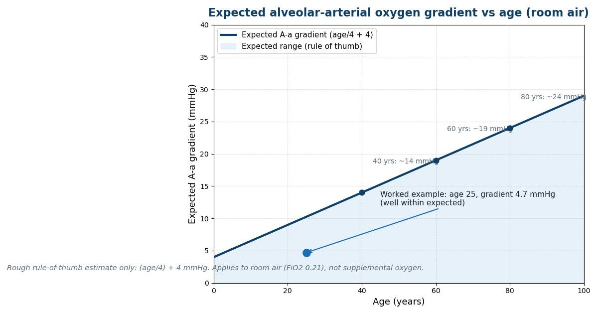
A gradient at or below the estimate is read as within the expected range; a gradient above it is read as widened, meaning gas exchange is impaired and the cause of hypoxemia lies in the lung itself rather than outside it. When the patient is on supplemental oxygen, the calculator deliberately withholds the age comparison: the gradient is physiologically larger on added oxygen (as the 163 mmHg example above shows), and applying a room-air rule of thumb would mislead. In that setting the number still has meaning, but it must be interpreted in clinical context, for example by watching how the gradient responds as FiO2 changes.
What widens the gradient: V/Q mismatch
The most common reason for a widened gradient is ventilation-perfusion mismatch. V/Q mismatch means some lung units get ventilation without matching blood flow (wasted ventilation, or dead space) while others get blood flow without matching ventilation (wasted perfusion, which behaves like a partial shunt). In low V/Q regions, blood leaves the alveoli poorly oxygenated, and because the oxygen-haemoglobin dissociation curve is flat at the top, the well-ventilated regions cannot add enough extra oxygen to compensate: haemoglobin there is already nearly saturated, so extra alveolar oxygen adds only a small amount of dissolved oxygen. The result is arterial blood that carries less oxygen than the average alveolar gas would predict, and the gradient widens. Typical causes include asthma, chronic obstructive pulmonary disease, pulmonary embolism, and pneumonia: all distort the matching of air and blood across the lung.
V/Q mismatch has one clinically useful property: it improves when the inspired oxygen is raised. Giving more oxygen to the poorly ventilated units raises their alveolar oxygen enough to load haemoglobin fully, so the gradient narrows and the PaO2 rises. That response is exactly what the 100 percent oxygen test exploits: if the hypoxemia corrects on high FiO2, the mechanism is likely V/Q mismatch rather than true shunt.
Shunt: why the gradient stays wide on 100 percent oxygen
True right-to-left shunt is blood that reaches the arterial side without ever seeing ventilated alveoli: blood flowing past collapsed or fluid-filled alveoli, or blood bypassing the lungs entirely through a cardiac defect. Shunted blood is venous blood, and no amount of extra oxygen in the alveoli can reach it, because it never passes through a ventilated unit. That is why shunt is the one cause of hypoxemia that does not correct on 100 percent oxygen: the gradient stays wide even at FiO2 1.0. In practice the calculator flags this pattern when a very wide gradient persists on high FiO2 with ongoing hypoxemia. Common clinical settings include acute respiratory distress syndrome, where alveoli flood with inflammatory fluid; large pneumonias that consolidate whole lobes; and atelectasis, where alveoli collapse. Quantifying shunt formally requires mixed venous sampling and a shunt equation, which this calculator does not attempt; the gradient on 100 percent oxygen is used here only as a pointer toward the possibility, to be confirmed by clinical assessment.
Diffusion limitation
The third classic cause is diffusion limitation: oxygen crosses the alveolar-capillary membrane too slowly for blood to equilibrate during the time the red cell spends in the capillary. At rest the transit time is generous and diffusion limitation rarely matters; during exercise, when cardiac output rises and transit time shortens, it can appear. Diseases that thicken or destroy the membrane, such as pulmonary fibrosis and emphysema, widen the gradient by this route. Like V/Q mismatch, diffusion limitation improves with added oxygen, because a larger driving gradient pushes more oxygen across the membrane per unit time.
When the gradient stays normal despite low oxygen
A low PaO2 with a normal A-a gradient is just as informative as a widened one: it says the lung is exchanging gas normally and the problem is elsewhere. The classic example is hypoventilation. When alveolar ventilation falls, carbon dioxide accumulates (PaCO2 rises) and, by the alveolar gas equation, alveolar oxygen falls by the same arithmetic. Arterial oxygen follows it down by an equal amount, so the gap between them stays normal. The calculator flags this pattern: raised PaCO2 with a normal gradient points toward hypoventilation as the driver of hypoxemia, for example in opioid-induced respiratory depression, neuromuscular weakness, or severe obesity hypoventilation. The second classic cause is low inspired oxygen: at altitude the barometric pressure falls, so (Patm minus 47) times FiO2 falls even though FiO2 is still 0.21, and both PAO2 and PaO2 fall together with a normal gradient. The calculator handles this through the barometric pressure input, which is why it is adjustable rather than fixed at 760. A third setting is simply breathing a hypoxic gas mixture, as in some industrial exposures. In all three, the lung itself is doing its job; fixing the hypoxemia means fixing ventilation or the inspired oxygen, not the lung.
Limitations and pitfalls
Several assumptions can make the number misleading, and each is worth checking before acting on the result. First, the respiratory quotient is a guess: 0.8 is the steady-state average for a typical mixed diet, but it moves toward 1.0 on a carbohydrate-heavy diet and toward 0.7 during starvation or prolonged fasting. In most patients the error this introduces is a few millimetres of mercury, but in critically ill or malnourished patients it is larger. Second, the PaO2 must come from arterial blood. A venous sample, or an arterial sample diluted with venous blood from a difficult draw, will report a falsely low PaO2 and a falsely wide gradient. Third, pulse oximetry is not a substitute: SpO2 measures haemoglobin saturation, not dissolved oxygen tension, and the flat top of the dissociation curve means very different PaO2 values can share the same saturation. Fourth, a negative gradient (calculated PAO2 below the measured PaO2) is not physiology; it signals an error in the inputs, the FiO2, or the sample, and the calculator flags it as such. Fifth, temperature matters: the 47 mmHg water vapour constant assumes body temperature of 37 C, and severe hypothermia or fever shifts it slightly. Finally, the age estimate is a rule of thumb derived for room air at sea level in otherwise healthy people; it is a guide for interpretation, not a diagnosis, and it should never override clinical judgment or replace arterial blood gas analysis interpreted by a clinician.
When clinicians use this calculation
The gradient earns its place in the workup of unexplained hypoxemia. When a patient is hypoxemic and the cause is not obvious, computing the gradient sorts the possibilities in seconds: widened means look at the lung (V/Q mismatch, shunt, diffusion block), normal means look outside it (ventilation, inspired oxygen). It is a routine part of interpreting arterial blood gases in emergency departments, intensive care units, and perioperative care, and it is embedded in the assessment of acute respiratory distress, where a wide gradient on high oxygen raises concern for shunt physiology. It also helps judge whether supplemental oxygen is doing what it should: if the gradient narrows as FiO2 rises, gas exchange is responding; if it stays wide, the mechanism resists oxygen therapy and the diagnostic focus shifts. For students, the calculation is the bridge between the alveolar gas equation on paper and the blood gas in hand; for clinicians, it is one of the fastest ways to turn a confusing low PaO2 into a short, testable differential. Like every number on this site, it is an educational aid, not a substitute for clinical assessment.
Sources
Physiology and reference values verified against: (1) StatPearls, "Physiology, Alveolar to Arterial Oxygen Gradient" (A-a gradient = PAO2 minus PaO2; alveolar gas equation PAO2 = (Patm minus PH2O) times FiO2 minus PaCO2/RQ; expected gradient estimated by (Age + 10)/4), https://www.ncbi.nlm.nih.gov/books/NBK545153/; (2) StatPearls, "Respiratory Failure in Adults" (FiO2 0.21 at room air, barometric pressure 760 mmHg, water vapour pressure 47 mmHg at 37 C, R 0.8 standard; normal A-a gradient with alveolar hypoventilation and low inspired oxygen), https://www.ncbi.nlm.nih.gov/books/NBK526127/. The age estimate (age/4) + 4 used by this calculator is a commonly cited rule-of-thumb form of the published estimator above, and is labelled as an estimate throughout.
Key takeaways
- There is no single universal cutoff, because the expected gradient rises with age.
- Even healthy lungs have a small physiologic ventilation-perfusion (V/Q) mismatch because blood flow and ventilation are distributed unevenly (the lung bases get relatively more perfusion, the apices relatively more ventilation).
- It means the lung is exchanging gas normally and the cause of the hypoxemia lies outside the alveolar-capillary membrane.
- No.
Frequently asked questions
What is a normal A-a gradient?
There is no single universal cutoff, because the expected gradient rises with age. A commonly cited rule of thumb estimates the expected gradient on room air as (age divided by 4) plus 4 mmHg: about 9 mmHg at age 20, 14 at age 40, and 19 at age 60. Treat this as a rough guide, not a diagnostic threshold, and apply it only on room air (FiO2 0.21): the gradient is physiologically much larger on supplemental oxygen.
Why does the expected A-a gradient rise with age?
Even healthy lungs have a small physiologic ventilation-perfusion (V/Q) mismatch because blood flow and ventilation are distributed unevenly (the lung bases get relatively more perfusion, the apices relatively more ventilation). That mismatch slowly increases as lungs age, so the gap between alveolar and arterial oxygen widens. The age estimate simply captures this trend.
My PaO2 is low but my A-a gradient is normal. What does that mean?
It means the lung is exchanging gas normally and the cause of the hypoxemia lies outside the alveolar-capillary membrane. The two classic causes are hypoventilation (raised PaCO2 lowers alveolar oxygen by the alveolar gas equation, and arterial oxygen falls by the same amount, so the gap stays normal) and low inspired oxygen, for example at altitude or when breathing a hypoxic gas mixture.
Can I use pulse oximetry (SpO2) instead of PaO2?
No. SpO2 measures haemoglobin oxygen saturation, not the dissolved oxygen tension in arterial blood. Because the oxygen-haemoglobin dissociation curve is flat at the top, very different PaO2 values can share the same saturation, so a gradient computed from SpO2 would be meaningless. Use PaO2 from an arterial blood gas.
Why is my A-a gradient still high while breathing 100 percent oxygen?
A gradient that stays wide on FiO2 1.0 points toward right-to-left shunt: blood reaching the arterial side without ever passing ventilated alveoli (for example collapsed or fluid-filled alveoli in ARDS or large pneumonias). V/Q mismatch and diffusion limitation improve with added oxygen, but shunted blood never sees the extra oxygen, so the hypoxemia does not correct. This is a pointer for clinical assessment, not a diagnosis.
Does altitude change the A-a gradient calculation?
Altitude changes the inputs, not the method. At altitude the barometric pressure is lower, so (Patm minus 47) times FiO2 gives a smaller alveolar oxygen even though FiO2 is still 0.21. Enter the local barometric pressure (the calculator defaults to 760 mmHg at sea level) and the equation handles the rest; PAO2 and PaO2 typically fall together with a normal gradient.
References and further reading
Medical disclaimer. This calculator is an educational tool, not medical advice. The A-a gradient must be interpreted together with the full clinical picture by a qualified clinician. Do not use it to diagnose, treat, or make decisions about any medical condition. If you or someone else has breathing difficulty, seek urgent medical care.