ABG Analyzer: Systematic Arterial Blood Gas Interpreter
In short: Free systematic ABG interpreter: enter pH, PaCO2 and HCO3 to identify the primary acid-base disorder, check compensation with Winter's formula and the 1-2-3-4-5 rule, and compute the albumin-corrected anion gap with delta gap and delta ratio. Worked examples and FAQs included. Use the calculator above, then read the guide below to interpret your result and its limitations.
Enter pH, PaCO2 and HCO3, with optional electrolytes, albumin, glucose and BUN. The analyzer names the primary acid-base disorder, checks whether compensation is appropriate using Winter's formula and the 1-2-3-4-5 rule, computes the albumin-corrected anion gap, and hunts for a second disorder with the delta gap and delta ratio. Medically reviewed by Dr. Taimoor Asghar. Formulas verified 2026-10-05 against the sources listed at the bottom of the page.
Analyze the blood gas
Required: pH, PaCO2 (mmHg), HCO3 (mEq/L). Optional: sodium, chloride and albumin unlock the anion gap; glucose and BUN add a calculated osmolarity.
Educational tool only: it applies standard interpretation rules to the numbers entered. It does not diagnose, and it cannot see the clinical context that often decides the final interpretation.
How to read an ABG in seven systematic steps
Arterial blood gas interpretation rewards routine. The same seven questions, asked in the same order, will untangle almost every gas you meet, from a straightforward diabetic ketoacidosis to a triple disorder in a ventilated patient. The analyzer above follows exactly this sequence, and this section explains each step so you can follow the machine's reasoning and catch it when the numbers do something unusual.
- Is the pH acidemic or alkalemic? Below 7.35 is acidemia; above 7.45 is alkalemia. This is the anchor of the whole interpretation, because the primary disorder is defined as the one that moves the pH in the observed direction.
- Which limb moves with the pH? If the pH is low and the bicarbonate is low, the primary disorder is a metabolic acidosis. If the pH is low and the PaCO2 is high, it is a respiratory acidosis. The same logic in reverse sorts out the alkaloses. When both limbs move in the pH's direction, the more deviant one is named primary and a mixed disorder is flagged.
- Is the compensation appropriate? The body defends pH automatically: the lungs compensate for metabolic disorders within minutes, and the kidneys compensate for respiratory disorders over days. Each primary disorder has an expected compensation, computed from a formula. A measured value outside the expected band means a second disorder is riding along.
- Calculate the anion gap, always. Even when the primary disorder looks respiratory, the gap can reveal a hidden metabolic acidosis. Correct it for albumin before judging it.
- If the gap is high, compute the delta gap and delta ratio. The rise in the gap should be matched by an equal fall in bicarbonate. Any mismatch exposes a co-existing normal anion gap acidosis or metabolic alkalosis.
- For respiratory disorders, judge acute versus chronic. Renal compensation takes 3 to 5 days to complete, so the size of the bicarbonate shift for a given PaCO2 change tells you how long the disorder has been running.
- Generate the differential. Once the disorders are named, the clinical question becomes why: the causes of each disorder are a separate diagnostic exercise, and the gas is only the map.
One rule governs the entire method and is worth stating plainly: physiologic compensation never normalizes the pH. If the pH sits comfortably between 7.35 and 7.45 while PaCO2 and bicarbonate are both abnormal, you are looking at a minimum of two primary disorders pulling in opposite directions. The analyzer flags this pattern explicitly.
pH first: acidemia, alkalemia, and the primary disorder
The pH cutoffs are among the least controversial numbers in medicine. Normal arterial pH is 7.35 to 7.45, corresponding to a hydrogen ion concentration of about 35 to 45 nmol/L. Below 7.35 is acidemia, above 7.45 is alkalemia. Note the careful words: acidemia and alkalemia describe the blood, while acidosis and alkalosis describe the processes. A patient can have a metabolic acidosis (the process) with a normal pH if a simultaneous metabolic alkalosis is cancelling it out. This is why step 1 looks at the blood and step 2 names the process.
Naming the primary disorder is a direction-matching exercise. Carbon dioxide is an acid: a high PaCO2 pushes pH down (respiratory acidosis) and a low PaCO2 pushes pH up (respiratory alkalosis). Bicarbonate is a base: a low bicarbonate pushes pH down (metabolic acidosis) and a high bicarbonate pushes pH up (metabolic alkalosis). When the pH is abnormal, the abnormality that points the same way as the pH is the primary disorder. The analyzer uses normal ranges of 35 to 45 mmHg for PaCO2 and 22 to 26 mEq/L for bicarbonate, and when both limbs point with the pH it compares their fractional deviations from normal to name the primary one, while flagging the case as possibly mixed.
A sanity check sits underneath all of this: the Henderson-Hasselbalch relationship. Given PaCO2 and bicarbonate, the pH is essentially fixed by chemistry: pH = 6.1 + log10(HCO3 / (0.03 x PaCO2)). If the measured pH disagrees with the calculated pH by more than a small margin, the numbers did not come from the same blood. The usual explanations are a venous sample mislabeled as arterial, a transcription error, or a sample left sitting long enough for ongoing metabolism to change it. The analyzer warns you when the three values are internally inconsistent, because no compensation formula can rescue a gas that contradicts its own chemistry.
Compensation formulas and where they come from
Compensation is the body's automatic counter-move, and each primary disorder has a predictable one. These are empirical rules derived from observing real patients, not laws of physics, so every formula carries a tolerance band. A measured value inside the band is appropriate compensation; a value outside it is a second primary disorder. The table below collects the formulas the analyzer uses, all traceable to standard references.
| Primary disorder | Expected compensation | Source |
|---|---|---|
| Metabolic acidosis | Expected PaCO2 = 1.5 x HCO3 + 8, plus or minus 2 mmHg (Winter's formula) | Winter et al.; RK.MD blood gas guide |
| Metabolic alkalosis | Expected PaCO2 = 0.7 x HCO3 + 21, plus or minus 2 mmHg | Standard acid-base references; LITFL |
| Acute respiratory acidosis | HCO3 rises 1 mEq/L for each 10 mmHg rise in PaCO2 (plus or minus 2) | 1-2-3-4-5 rule; LITFL |
| Chronic respiratory acidosis | HCO3 rises 3.5 mEq/L for each 10 mmHg rise in PaCO2 (plus or minus 2) | 1-2-3-4-5 rule; LITFL |
| Acute respiratory alkalosis | HCO3 falls 2 mEq/L for each 10 mmHg fall in PaCO2 (plus or minus 2) | 1-2-3-4-5 rule; LITFL |
| Chronic respiratory alkalosis | HCO3 falls 4 to 5 mEq/L for each 10 mmHg fall in PaCO2 (plus or minus 2) | 1-2-3-4-5 rule; LITFL |
Winter's formula deserves a closer look because it is the most-used equation in acid-base medicine. It describes the expected respiratory compensation for a metabolic acidosis: the patient hyperventilates, blowing off CO2, and the PaCO2 falls in proportion to the bicarbonate. The constant 8 and the slope 1.5 were fitted to patient data, and the plus-or-minus 2 mmHg band is part of the formula, not an afterthought. Two shortcuts are worth knowing for bedside use: the expected PaCO2 is roughly the last two digits of the pH (a pH of 7.15 predicts a PaCO2 near 15), and the PaCO2 falls about 1.2 mmHg for each 1 mEq/L fall in bicarbonate. The analyzer uses the full formula, but the shortcuts are handy for checking its answer.
The asymmetry of compensation timing matters. Respiratory compensation for a metabolic disorder is nearly immediate: chemoreceptors sense the pH change and ventilation adjusts within minutes. Renal compensation for a respiratory disorder takes 3 to 5 days, because the kidney must generate new bicarbonate by excreting ammonium and reclaiming filtered bicarbonate. This is why a respiratory acidosis with a bicarbonate that has only risen 1 per 10 mmHg is read as acute, while the same PaCO2 with a bicarbonate risen 3.5 per 10 mmHg is read as chronic. The chart on this page plots Winter's compensation band so you can see where a patient's values fall.
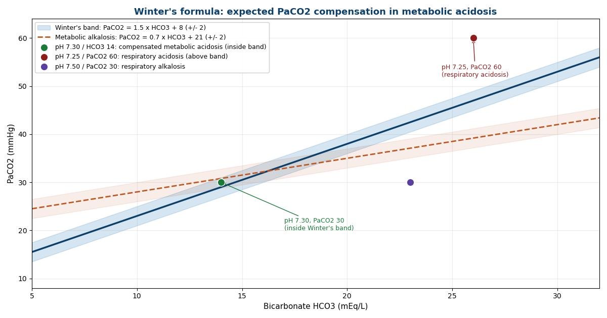
When the measured compensation misses the band, the miss has a name. In a metabolic acidosis, a PaCO2 above the expected band is a superimposed respiratory acidosis (the patient is not blowing off enough CO2, think fatigue, sedation, or lung disease), and a PaCO2 below the band is a superimposed respiratory alkalosis (think pain, anxiety, sepsis, or early salicylate toxicity driving extra ventilation). In a primary respiratory disorder, a bicarbonate higher than either the acute or chronic expectation is a superimposed metabolic alkalosis, and one lower than the acute expectation is a superimposed metabolic acidosis. The analyzer reports these misses in exactly these terms.
The anion gap, and why albumin correction is not optional
The anion gap quantifies the unmeasured anions in plasma: anion gap = Na minus (Cl + HCO3). The normal value is about 12 mEq/L, with a reference range of roughly 8 to 16. A gap above 16 (some sources use above 12 in the right clinical setting) indicates a high anion gap metabolic acidosis, meaning an acid has been added to the blood: lactate, ketoacids, uremic anions, or a toxin such as methanol, ethylene glycol, or salicylates. The classic teaching mnemonic for the causes is KULT: ketones, uremia, lactate, toxins.
The catch is that albumin is the largest contributor to the unmeasured anions, and sick patients are frequently hypoalbuminemic. Every 1 g/dL fall in albumin below the normal 4.0 g/dL lowers the anion gap by about 2.5 mEq/L, which can completely mask a significant high gap acidosis. The correction is simple: corrected gap = measured gap + 2.5 x (4.0 minus albumin). A patient with a measured gap of 14 and an albumin of 2.0 g/dL has a corrected gap of 19, which is a high gap metabolic acidosis that the uncorrected number would have missed. The analyzer applies this correction automatically whenever you enter an albumin below 4.0, and labels the result as albumin-corrected so the provenance is clear.
A low anion gap is less commonly useful but worth a mention: it usually reflects hypoalbuminemia (the same correction explains it), or occasionally an increase in unmeasured cations such as in multiple myeloma, lithium toxicity, or bromide ingestion, or a laboratory artifact from hyperlipidemia or hyperviscosity. The analyzer reports the gap with its reference range and lets the low values speak for themselves.
Delta gap and delta ratio: hunting the second disorder
Once a high anion gap metabolic acidosis is established, the next question is whether it is traveling alone. The logic is stoichiometric: each retained acid anion consumes one bicarbonate, so the rise in the anion gap (delta AG = AG minus 12) should be matched by an equal fall in bicarbonate (delta HCO3 = 24 minus HCO3). Two derived numbers exploit this one-to-one accounting.
The delta gap is the rise in the gap minus the fall in bicarbonate: delta gap = (AG minus 12) minus (24 minus HCO3). If it is near zero, the bicarbonate fell exactly as much as the gap rose and the high gap acidosis is the whole story. If it is positive, the bicarbonate is higher than the gap alone explains, which means a concurrent metabolic alkalosis (or a chronic respiratory acidosis) was already holding bicarbonate up. If it is negative, the bicarbonate fell further than the gap rose, which means a concurrent normal anion gap (hyperchloremic) acidosis consumed the extra bicarbonate. A practical shortcut: add the delta gap to the measured bicarbonate. A result above 30 suggests co-existing metabolic alkalosis; below 23 suggests a co-existing normal gap acidosis.
The delta ratio divides the rise in the gap by the fall in bicarbonate: delta ratio = (AG minus 12) / (24 minus HCO3). The standard interpretation bands, as published on Life in the Fast Lane's acid-base reference, are:
| Delta ratio | Interpretation |
|---|---|
| Below 0.4 | Pure normal anion gap (hyperchloremic) acidosis |
| 0.4 to 0.8 | Combined normal and high anion gap acidosis |
| 0.8 to 2.0 | Pure high anion gap metabolic acidosis |
| Above 2.0 | High anion gap acidosis plus metabolic alkalosis, or a pre-existing compensated respiratory acidosis |
Two caveats keep these numbers honest. First, the ratio is only meaningful when both the gap is elevated and the bicarbonate is below 24; dividing by a zero or negative bicarbonate fall produces nonsense, so the analyzer declines to compute it in that case and says why. Second, the ratio drifts in renal failure, where it often runs below 1 even in a pure high gap acidosis, because uremic anions distribute differently. The bands are guides for reasoning, not diagnostic cutoffs with legal force.
Mixed disorders you should learn to recognize
Mixed acid-base disorders are common in sick patients and are exactly what the compensation checks and delta calculations exist to find. A few classic patterns are worth memorizing. A high anion gap metabolic acidosis with a PaCO2 above Winter's expectation is a metabolic acidosis plus a respiratory acidosis: think a shocked patient tiring out, or an ingestion with central respiratory depression. The same metabolic acidosis with a PaCO2 below expectation is a metabolic acidosis plus a respiratory alkalosis: think sepsis, salicylate toxicity (which classically produces a respiratory alkalosis plus a high gap metabolic acidosis), or liver failure.
Vomiting with volume depletion produces a metabolic alkalosis, and if the patient is also in ketoacidosis or lactic acidosis, the delta ratio will climb above 2 as the retained bicarbonate from vomiting offsets the bicarbonate consumed by the acids. Diarrhea produces a normal gap acidosis, so a high gap acidosis with a delta ratio below 0.8 is often a lactic acidosis plus bicarbonate loss from the gut or the kidney. And the famous normal-pH trap: a pH of 7.40 with a PaCO2 of 60 and a bicarbonate of 36 is not a normal gas. It is a respiratory acidosis and a metabolic alkalosis in perfect opposition, and treating only one of them will unmask the other.
The analyzer reports each of these patterns in plain language when the numbers fit. It does not know the patient, so it phrases findings as possibilities to check rather than diagnoses to act on: "measured PaCO2 above the expected band suggests an additional respiratory acidosis" rather than "the patient has respiratory acidosis." That phrasing is deliberate.
Worked examples
Example 1: pH 7.25, PaCO2 60, HCO3 26. The pH is acidemic. Bicarbonate is at the upper edge of normal and does not move with the pH; PaCO2 is high and does move with it, so the primary disorder is a respiratory acidosis. For chronicity, a PaCO2 rise of 20 mmHg predicts a bicarbonate of 26 in the acute pattern (24 + 1 x 2) and 31 in the chronic pattern (24 + 3.5 x 2). The measured 26 matches the acute expectation, so this reads as an acute respiratory acidosis, for example early in an asthma exacerbation or after oversedation, before the kidneys have had days to compensate.
Example 2: pH 7.30, PaCO2 30, HCO3 14. Acidemia with a low bicarbonate: primary metabolic acidosis. Winter's formula predicts a PaCO2 of 1.5 x 14 + 8 = 29, plus or minus 2, giving an expected band of 27 to 31. The measured 30 sits inside the band, so compensation is appropriate: this is a compensated metabolic acidosis with no second respiratory disorder. The low PaCO2 here is the body's correct response, not a separate alkalosis.
Example 3: pH 7.50, PaCO2 30, HCO3 23. Alkalemia with a low PaCO2: primary respiratory alkalosis. The PaCO2 has fallen 10 mmHg, so the acute expectation is a bicarbonate of 22 (24 minus 2) and the chronic expectation is about 19.5 (24 minus 4.5), each plus or minus 2. The measured 23 fits the acute pattern: an acute respiratory alkalosis with appropriate compensation, as seen in acute anxiety-driven hyperventilation or early sepsis.
Limitations and pitfalls
Every rule on this page has boundary conditions. Winter's formula and the respiratory compensation rules were derived from otherwise stable patients; they become unreliable at extremes, in rapidly changing situations, and when the bicarbonate is very low or very high. The formulas assume standard units (mmHg and mEq/L); entering kPa or mmol/L values from a non-US analyzer without converting will produce confident nonsense, which is why the analyzer rejects values outside plausible ranges with named errors rather than computing silently.
The albumin correction assumes the gap's only moving part is albumin, which is usually but not always true. The delta ratio bands assume a baseline gap of 12 and a baseline bicarbonate of 24, which do not hold for every patient; someone whose baseline bicarbonate was 30 before they became ill will generate a misleading delta gap. Chronic respiratory disorders need history to date them: the bicarbonate expectations overlap in the first days, and no formula can tell day two from day four. Finally, venous blood gases differ systematically from arterial ones (venous pH runs about 0.03 to 0.05 lower and PCO2 about 4 to 5 mmHg higher), so a venous sample run through arterial rules will look like a mild respiratory acidosis that is not really there. When in doubt, the sample source matters more than the arithmetic.
Sources and verification
The compensation formulas and interpretation bands on this page were verified on 2026-10-05 against: Life in the Fast Lane: Acid-base disorders (Winter's rule, the 1-2-3-4-5 rule for acute and chronic respiratory compensation, and the delta ratio interpretation bands); RK.MD: Arterial Blood Gas (ABG) Interpretation (Winter's formula as PaCO2 expected = 1.5 x HCO3 + 8 plus or minus 2, and the delta/delta 1.0 to 2.0 range for pure high anion gap metabolic acidosis); and the Acid Base 101 reference sheet (albumin correction of 2.5 mEq/L per 1 g/dL below 4 g/dL). No statistics, quotations, or thresholds on this page come from uncited sources.
Key takeaways
- Look at the pH before anything else.
- Winter's formula estimates the expected respiratory compensation in metabolic acidosis: expected PaCO2 = 1.5 x HCO3 + 8, plus or minus 2 mmHg.
- Albumin is the main unmeasured anion, so a low albumin artificially lowers the anion gap and can hide a high anion gap metabolic acidosis.
- In a high anion gap metabolic acidosis, the rise in the gap should be matched by an equal fall in bicarbonate, because each retained acid anion consumes one bicarbonate.
Frequently asked questions
What is the first step when interpreting an arterial blood gas?
Look at the pH before anything else. A pH below 7.35 is acidemia and a pH above 7.45 is alkalemia, and the primary disorder is the one that moves the pH in that direction. Only after naming the primary disorder do you check whether the other limb (PaCO2 or bicarbonate) is compensating appropriately, because compensation never fully normalizes the pH: a normal pH with abnormal values means at least two disorders are present.
What is Winter's formula and when is it used?
Winter's formula estimates the expected respiratory compensation in metabolic acidosis: expected PaCO2 = 1.5 x HCO3 + 8, plus or minus 2 mmHg. It is used only when the primary disorder is a metabolic acidosis. If the measured PaCO2 is higher than expected, a superimposed respiratory acidosis is present; if it is lower, a superimposed respiratory alkalosis is present. Respiratory compensation for metabolic disorders is nearly immediate, so the formula applies at presentation.
Why does the anion gap need an albumin correction?
Albumin is the main unmeasured anion, so a low albumin artificially lowers the anion gap and can hide a high anion gap metabolic acidosis. The standard correction adds 2.5 mEq/L to the gap for every 1 g/dL that albumin falls below the normal value of 4.0 g/dL. In a critically ill patient with an albumin of 2.0 g/dL, the gap is corrected upward by 5 mEq/L, which is often the difference between a missed and a caught diagnosis.
What do the delta gap and delta ratio tell you?
In a high anion gap metabolic acidosis, the rise in the gap should be matched by an equal fall in bicarbonate, because each retained acid anion consumes one bicarbonate. The delta gap is the rise in the gap minus the fall in bicarbonate: a positive value means bicarbonate is higher than expected (a co-existing metabolic alkalosis), a negative value means it is lower (a co-existing normal anion gap acidosis). The delta ratio divides the rise in the gap by the fall in bicarbonate: below 0.4 suggests pure normal anion gap acidosis, 0.4 to 0.8 a mixed high and normal gap acidosis, 0.8 to 2.0 a pure high gap acidosis, and above 2.0 a high gap acidosis plus metabolic alkalosis or chronic respiratory acidosis.
How do you tell acute from chronic respiratory disorders on one blood gas?
By how far the bicarbonate has moved for the change in PaCO2, using the 1-2-3-4-5 rule. In respiratory acidosis, bicarbonate rises about 1 mEq/L per 10 mmHg rise in PaCO2 when acute and about 3.5 per 10 mmHg when chronic. In respiratory alkalosis, bicarbonate falls about 2 per 10 mmHg when acute and 4 to 5 per 10 mmHg when chronic. Matching the measured bicarbonate against both expectations shows which pattern fits; clinical history remains the final arbiter because renal compensation takes days to develop fully.
Can this analyzer replace clinical judgment or diagnose a patient?
No. It is an educational tool that applies standard textbook rules to the numbers you enter. It cannot know the clinical context that often decides the interpretation, such as how long a respiratory disorder has been present, whether the sample was truly arterial, or what the baseline values were. Severe or rapidly changing acid-base disturbances need urgent in-person assessment, and treatment decisions belong to the treating clinician.
Related pulmonology tools
References and further reading
Last medically reviewed 2026-10-05 by Dr. Taimoor Asghar. Formulas and interpretation bands verified against the cited sources on 2026-10-05.